What is the difference between Types of Neuromodulation?
All the new neuromodulation modalities can be confusing to tell apart. How are they all different, what is their history, and how can they help you.
Did you enjoy this article? Checkout the podcast here: https://gettherapybirmingham.podbean.com/

What is qEEG Brain Mapping?
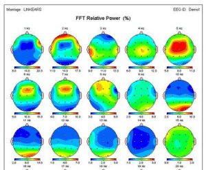
Quantitative Electroencephalogram (qEEG) Brain Mapping is a valuable diagnostic test used to evaluate patients with various neurological and psychological conditions. qEEG Brain Mapping provides a non-invasive way for clinicians to analyze the functioning of the brain and identify the source of symptoms or dysregulation. By establishing maximally effective neurofeedback treatment protocols, patients can improve their quality of life without relying on medication.
During a qEEG Brain Mapping session, 19 sensors are placed on the surface of the head, and brain wave activity is recorded over those 19 areas. The test is non-invasive, painless, and records only the electrical activity of the brain for analysis. Brain map recordings are processed through an FDA-approved normative database for comparison to “normal” results, which allows for the identification of any abnormal areas that require specific attention during neurofeedback treatment.
The qEEG Brain Mapping process is comparable to a physician doing a bacterial culture to determine which antibiotic would best treat an infection. Similarly, the brain map tells us if the symptoms are neurologically based or linked. When symptoms are neurologically linked, there is a high probability of treatment success using neurofeedback.
The qEEG Brain Mapping process detects if any area of the brain is dysfunctional or dysregulated. Sometimes, symptoms are caused by an area or areas of the brain that are underactive, showing excessive slow brain waves that cause impaired functioning and symptoms. Conversely, a smaller percentage of patients will exhibit symptoms due to an area or areas of his/her brain that are overstimulated, showing too many fast brain waves. Either abnormality is disruptive to normal or optimal functioning and can cause clinical symptoms.
Research in the neurosciences has shown that functional connections between brain regions are key for optimal brain functioning. The qEEG brain map analysis measures “Coherence” between different areas, which indicates the health or normalcy of functional communications between these regions of the brain; in other words, it measures how well the different areas of the brain are communicating with each other to perform complex tasks.
qEEG Brain Mapping is particularly useful for patients with ADHD and Autism Spectrum Disorder. These conditions often result from a misfiring or dysregulation within a specific brain network or the failure of networks to work together to carry out complex mental functioning or behavioral/emotional control. The test helps identify the specific source of the symptoms and provides insights into which treatment protocols will be most effective.
What Does qEEG Brain Mapping Tell You?
The analysis of your qEEG brain map provides us with valuable information about how your brain functions and how it may contribute to your symptoms and challenges. We use this information to design your individualized therapy plan.
The written summary of your brain maps connects your brain function to your symptoms, cognitive issues, and behavioral challenges. We integrate brain mapping into a comprehensive assessment, which guides us in developing your individualized therapy plan. By utilizing this information, we can create a tailored treatment approach that addresses the root causes of your challenges and enhances your therapy outcomes.
How Can qEEG Brain Mapping Help You?

qEEG brain mapping can help identify areas of your brain that may not be functioning optimally and contributing to your symptoms and challenges. By understanding how your brain functions, we can create a personalized therapy plan that addresses the root causes of your challenges and enhances your therapy outcomes.
The qEEG brain map provides us with a valuable tool to monitor your brainwave changes and progress with repeated qEEG and other assessments as needed. This process helps us evaluate the effectiveness of your therapy and make necessary adjustments to your treatment plan.
At our psychotherapy practice, we are committed to providing our clients with the highest quality of care. By utilizing cutting-edge techniques like qEEG brain mapping, we can enhance our therapy approach and improve your therapy outcomes.
In conclusion, qEEG brain mapping is a non-invasive and effective technique that can provide valuable insights into how your brain functions and how it may contribute to your symptoms and challenges. By incorporating this technique into our therapy approach, we can design a personalized treatment plan that addresses the root causes of your challenges and enhances your therapy outcomes. Contact us today to learn more about our therapy services and how qEEG brain mapping can inform your treatment process.
What does qEEG Brain Mapping Detect? What Kinds of thinking can it change?
There are several types of brain waves that are typically analyzed, including delta, alpha, theta, beta, and high beta waves. Each of these brain waves is associated with different frequencies and has its own unique meaning when it comes to qEEG brain mapping.
Delta Waves:
Delta waves are the slowest type of brain waves, with a frequency of 0.5-4 Hz. These waves are typically associated with deep sleep and are also present when a person is in a coma. Abnormalities in delta waves can be associated with certain neurological conditions, such as traumatic brain injury, stroke, and dementia.
Alpha Waves:
Alpha waves have a frequency of 8-12 Hz and are typically present when a person is awake but relaxed. They are often observed when someone is closing their eyes or engaging in a meditative state. A decrease in alpha waves may be associated with anxiety or depression, while an increase in alpha waves may be associated with improved relaxation and stress reduction.
Theta Waves:
Theta waves have a frequency of 4-8 Hz and are typically observed during light sleep or drowsiness. They may also be present when a person is in a meditative or creative state. In qEEG brain mapping, an increase in theta waves may be associated with attention deficit hyperactivity disorder (ADHD), while a decrease in theta waves may be associated with cognitive decline in older adults.
Beta Waves:
Beta waves have a frequency of 12-30 Hz and are typically present when a person is awake and engaged in cognitive or physical activity. They are often associated with alertness, focus, and concentration. Abnormalities in beta waves can be associated with conditions such as anxiety, depression, and insomnia.
High Beta Waves:
High beta waves have a frequency of 30-40 Hz and are often associated with intense cognitive or physical activity, such as problem-solving or exercise. In qEEG brain mapping, an increase in high beta waves may be associated with conditions such as ADHD or obsessive-compulsive disorder (OCD).
Overall, the analysis of delta, alpha, theta, beta, and high beta waves in qEEG brain mapping can provide valuable information about a person’s neurological functioning and potential cognitive or mental health issues. By identifying abnormalities in these brain waves, healthcare professionals can develop more targeted and effective treatment plans for their patients.
What is Neurostimulation?
Neurostimulation is a field of medicine that involves the use of electrical or magnetic stimulation to treat a variety of neurological disorders. This technology has been used for decades to treat conditions such as chronic pain, depression, and Parkinson’s disease, and has undergone significant advancements in recent years.
History of Neurostimulation:
The earliest form of neurostimulation dates back to the ancient Greeks, who used electric fish to treat headaches and other ailments. In the 18th and 19th centuries, experiments with electricity and the nervous system became more widespread, and by the 20th century, neurostimulation was being used in clinical settings to treat chronic pain.
In the 1960s, the development of implantable electrodes allowed for more precise and targeted neurostimulation, and the first implantable spinal cord stimulator was introduced in 1967. This device, which delivered electrical impulses to the spinal cord, provided relief for chronic pain sufferers.
In the 1980s, deep brain stimulation (DBS) was introduced as a treatment for Parkinson’s disease. DBS involves implanting electrodes deep in the brain and delivering electrical impulses to specific areas to improve motor function and reduce tremors.
Advancements in Neurostimulation:
Since the introduction of deep brain stimulation (DBS), neurostimulation technology has continued to evolve. In the 1990s, transcranial magnetic stimulation (TMS) was introduced as a non-invasive alternative to DBS. TMS uses a magnetic field to stimulate the brain and has been shown to be effective in treating depression.
In the early 2000s, the first non-invasive spinal cord stimulator was introduced, allowing for targeted pain relief without surgery. And in recent years, advancements in technology have led to the development of closed-loop systems, which use sensors to monitor brain activity and adjust stimulation in real-time.
Today, neurostimulation is used to treat a wide range of conditions, including chronic pain, epilepsy, depression, and obsessive-compulsive disorder. As the technology continues to evolve, it is likely that even more conditions will be treated using neurostimulation.
How can Neurostimulation and QEEG Brain Mapping Change the Way You Think?
Neurostimulation and QEEG brain mapping are two technologies that can work together to open new pathways in the brain. Neurostimulation refers to the use of electrical or magnetic fields to stimulate nerves and alter brain activity. QEEG brain mapping, on the other hand, is a type of brain scan that uses electrodes placed on the scalp to measure brain waves and identify areas of abnormal activity.
Through the use of neurostimulation and QEEG brain mapping, it is possible to create new connections between different areas of the brain. For example, if QEEG brain mapping shows that there is reduced activity in a particular area of the brain, neurostimulation can be used to stimulate that area and increase its activity. This can lead to the creation of new neural pathways, which can improve cognitive functioning and enhance brain plasticity.
Neurostimulation and QEEG can also be used together to treat a variety of neurological and psychiatric conditions, including depression, anxiety, ADHD, and traumatic brain injury. By using neurostimulation to stimulate areas of the brain that are underactive or overactive, and by monitoring changes in brain activity with QEEG, it is possible to create individualized treatment plans that target specific areas of the brain.
In addition, neurostimulation and QEEG can be used to enhance cognitive performance in healthy individuals. For example, athletes may use neurostimulation and QEEG to improve their reaction times, while students may use these technologies to enhance their memory and concentration.
Overall, the combination of neurostimulation and QEEG brain mapping offers a powerful tool for opening new pathways in the brain and improving cognitive functioning. As these technologies continue to evolve, they are likely to play an increasingly important role in the treatment of neurological and psychiatric conditions, as well as in enhancing cognitive performance in healthy individuals.
What is Neurofeedback?
Neurofeedback therapy, also called EEG Biofeedback, is a non-invasive, drug-free method for training the brain to function in a more balanced and healthy way. It involves monitoring the electrical impulses in the brain using sensors and helping the brain learn to self-regulate by rewarding optimal brainwaves with bright visuals and sound. Neurofeedback can help people suffering from trauma, addiction, anxiety, depression, ADHD, autism, learning disorders, migraines, insomnia, memory loss, and those seeking peak performance in academics, sports, or other physical and intellectual pursuits.
Studies have shown that neurofeedback therapy is a safe and broadly effective treatment for people of all ages. In a randomized control trial with adolescents who had endured developmental trauma, researchers found that the use of neurofeedback therapy had a significant positive impact, resulting in an eleven times greater improvement in self-control and better emotional regulation capabilities compared to the control group.
Neurofeedback therapy can be done in-office or with a home system for convenience, and it offers an innovative drug-free alternative for those who have seen little to no improvement with medication. By teaching clients to self-regulate through operant conditioning, neurofeedback therapy helps them reach their full potential without any side effects.
What is Biofeedback?
Biofeedback therapy is a non-invasive form of therapy that involves using sensors to measure bodily functions such as heart rate, blood pressure, muscle tension, and skin temperature. These measurements are then displayed on a monitor, allowing patients to become more aware of their physiological responses and learn how to control them.
During a biofeedback session, a therapist attaches sensors to the patient’s skin, typically on the fingers or scalp, which measure physiological responses such as muscle tension or brain activity. The patient then receives visual or auditory feedback on a computer monitor, which provides real-time information on their physiological state. By learning to interpret and control these signals, patients can develop greater control over their bodily functions.
Biofeedback therapy has been used to treat a variety of conditions, including anxiety, chronic pain, headaches, high blood pressure, and digestive disorders. By learning to control their physiological responses, patients can reduce symptoms associated with these conditions and improve their overall quality of life.
One of the key benefits of biofeedback therapy is that it empowers patients to take an active role in their own treatment. By learning to control their physiological responses, patients can reduce their dependence on medication and other forms of intervention. Biofeedback therapy is also non-invasive and generally free from side effects, making it a safe and effective form of treatment for many individuals.
How can Brain Training and Biofeedback Change Cognition?
Biofeedback visual stimulation and brain training are two techniques that can be used to help improve cognitive function and reduce symptoms of various neurological disorders.
Biofeedback visual stimulation involves using visual cues, such as lights or patterns, to train the brain to respond in certain ways. This technique can be used to help with a variety of conditions, such as ADHD, anxiety, and chronic pain. The goal is to help the brain learn how to regulate its own activity in response to stimuli, which can lead to improved function and reduced symptoms.
Brain training, on the other hand, involves a range of techniques designed to improve cognitive function and performance. This can include exercises and games that challenge the brain in various ways, such as memory, attention, and problem-solving. Brain training can be used to improve cognitive function in healthy individuals, as well as those with neurological disorders.
Both biofeedback visual stimulation and brain training can be used in conjunction with other treatments, such as medication and therapy, to provide a comprehensive approach to managing neurological conditions. These techniques have been shown to be effective in improving cognitive function and reducing symptoms in a variety of conditions, and may offer new pathways for treatment and management of these conditions in the future.
What is Trans Cranial Magnetic Stimulation?
Transcranial magnetic stimulation (TMS) is a non-invasive procedure that uses magnetic fields to stimulate nerve cells in the brain to improve symptoms of major depression. The US FDA has also approved TMS for treating obsessive-compulsive disorder, migraines, and to help people quit smoking when standard treatments haven’t worked well. During an rTMS session, an electromagnetic coil is placed against the scalp of the head and delivers magnetic pulses that stimulate nerve cells in the brain region involved in mood control and depression. TMS seems to ease depression symptoms and improve mood by affecting how the brain is working. TMS is generally well-tolerated and the most common side effect is headache during or after treatment. Approximately 50-60% of people with depression who have tried and failed to receive benefit from medications experience a clinically meaningful response with TMS, with about one-third of these individuals experiencing a full remission. TMS therapy requires sessions that occur five days a week for several weeks, and each session may last anywhere from 20 to 50 minutes. TMS is also being studied across disorders and disciplines with the hope that it will evolve into new treatments for neurological disorders, pain management, and physical rehabilitation.
If you pre register for neurostim before Peak Neuroscience opens at Taproot Therapy Collective you will receive $50 off the first 10 appointments.
Learn more about neurostim or book an appointment.
Bibliography:
Ashton, M. (2023). What is the Difference Between Types of Neuromodulation? GetTherapyBirmingham.com.
Demos, J. N. (2005). Getting Started with Neurofeedback. W.W. Norton & Company.
Lefaucheur, J. P., Antal, A., Ayache, S. S., Benninger, D. H., Brunelin, J., Cogiamanian, F., … & Paulus, W. (2017). Evidence-Based Guidelines on the Therapeutic Use of Transcranial Direct Current Stimulation (tDCS). Clinical Neurophysiology, 128(1), 56-92. https://doi.org/10.1016/j.clinph.2016.10.087
Lipov, E., & Kelzenberg, B. (2012). Stellate Ganglion Block May Promote Neuroplasticity in PTSD: A Review of the Evidence. Journal of Trauma & Treatment, 1(4), 1-4. https://doi.org/10.4172/2167-1222.1000149
Megumi, F., Yamashita, A., Kawato, M., & Imamizu, H. (2015). Functional MRI Neurofeedback Training on Connectivity Between Two Regions Induces Long-Lasting Changes in Intrinsic Functional Network. Frontiers in Human Neuroscience, 9, 160. https://doi.org/10.3389/fnhum.2015.00160
Schabus, M., Griessenberger, H., Gnjezda, M. T., Heib, D. P., Wislowska, M., & Hoedlmoser, K. (2017). Better than Sham? A Double-Blind Placebo-Controlled Neurofeedback Study in Primary Insomnia. Brain, 140(4), 1041-1052. https://doi.org/10.1093/brain/awx011
Thibault, R. T., Lifshitz, M., & Raz, A. (2016). The Self-Regulating Brain and Neurofeedback: Experimental Science and Clinical Promise. Cortex, 74, 247-261. https://doi.org/10.1016/j.cortex.2015.10.024
Further Reading:
Arns, M., De Ridder, S., Strehl, U., Breteler, M., & Coenen, A. (2009). Efficacy of Neurofeedback Treatment in ADHD: the Effects on Inattention, Impulsivity and Hyperactivity: a Meta-Analysis. Clinical EEG and Neuroscience, 40(3), 180-189. https://doi.org/10.1177/155005940904000311
Fisher, C. E., Chin, L., & Klitzman, R. (2010). Defining Neuromarketing: Practices and Professional Challenges. Harvard Review of Psychiatry, 18(4), 230-237. https://doi.org/10.3109/10673229.2010.501497
Linden, D. E. (2014). Neurofeedback and Networks of Depression. Dialogues in Clinical Neuroscience, 16(1), 103.
Ros, T., Baars, B. J., Lanius, R. A., & Vuilleumier, P. (2014). Tuning Pathological Brain Oscillations with Neurofeedback: A Systems Neuroscience Framework. Frontiers in Human Neuroscience, 8, 1008. https://doi.org/10.3389/fnhum.2014.01008
Rubin, L. H., Witkiewitz, K., St. Andre, J., & Reilly, S. (2007). Methods for Mediation Analysis in Randomized Clinical Trials. Addiction, 102(11), 1785-1792. https://doi.org/10.1111/j.1360-0443.2007.01882.x


0 Comments