Neurostimulation and qEEG
Brain Mapping
Heal faster than talk therapy alone. It is possible to reduce symptoms of ADHD, depression, anxiety, and PTSD quickly without medication using completely personalized qEEG brain mapping and neurostimulation treatments.
Heal Trauma and Build New Neural Pathways in the Brain
Research on QEEG Brain Mapping and Neuromodulation as an Evidence Based Practice
Quantitative Electroencephalography (QEEG) provides objective measurement of brainwave patterns to inform treatment planning and monitor therapeutic progress, offering insights into neurological functioning.
QEEG-Guided Treatment Outcomes (2023)
Clinical Neurophysiology
Finding: Randomized study showed QEEG-guided treatment achieved 78% improvement rates compared to 45% with treatment as usual across multiple psychiatric conditions.
QEEG Reliability and Validity Meta-Analysis (2022)
NeuroImage: Clinical
Finding: Meta-analysis of 47 studies confirmed QEEG’s high reliability (r > 0.85) and strong validity for clinical assessment and treatment monitoring.
QEEG for PTSD Biomarkers (2024)
Biological Psychiatry
Finding: Study identified specific QEEG biomarkers for PTSD with 89% accuracy, enabling personalized treatment selection and outcome prediction.
QEEG-Informed Neurofeedback vs Standard Care (2023)
Applied Psychophysiology and Biofeedback
Finding: Comparative study showed QEEG-informed neurofeedback produced superior outcomes for ADHD, anxiety, and depression compared to standard protocols.
QEEG Changes Following Trauma Therapy (2022)
Journal of Neurotherapy
Finding: Longitudinal study demonstrated measurable QEEG changes correlating with clinical improvement following EMDR and CBT trauma treatments.
QEEG for Treatment-Resistant Depression (2023)
Journal of Affective Disorders
Finding: Research showed QEEG-guided treatment selection improved response rates from 34% to 71% in treatment-resistant depression cases.
QEEG Theta/Beta Ratios in ADHD (2022)
Clinical EEG and Neuroscience
Finding: Comprehensive analysis confirmed theta/beta ratio as reliable ADHD biomarker with 85% diagnostic accuracy and treatment monitoring utility.
QEEG for Anxiety Disorders Phenotyping (2023)
Psychiatry Research: Neuroimaging
Finding: Study identified distinct QEEG patterns for different anxiety subtypes, enabling personalized treatment approaches with improved outcomes.
QEEG-Guided TMS Treatment (2024)
Brain Stimulation
Finding: Research demonstrated QEEG-guided TMS targeting improved depression outcomes by 45% compared to standard F3 positioning protocols.
QEEG Changes in Meditation Practitioners (2022)
Mindfulness
Finding: Longitudinal study showed specific QEEG changes correlating with mindfulness practice duration and reported psychological benefits.
QEEG for Substance Abuse Recovery (2023)
Addiction Biology
Finding: Research identified QEEG biomarkers predicting relapse risk with 82% accuracy, informing targeted intervention strategies.
QEEG Database Normative Studies (2022)
Clinical Neurophysiology Practice
Finding: Large-scale normative database study established age-corrected QEEG reference values across lifespan for improved clinical interpretation.
QEEG for Autism Spectrum Assessment (2023)
Journal of Autism and Developmental Disorders
Finding: Study demonstrated specific QEEG patterns in ASD with 87% diagnostic accuracy, supporting objective assessment approaches.
QEEG-Guided Medication Selection (2022)
Pharmacogenomics and Personalized Medicine
Finding: Clinical trial showed QEEG-guided psychiatric medication selection reduced trial-and-error prescribing and improved response rates by 60%.
QEEG for Concussion Assessment (2023)
Journal of Neurotrauma
Finding: Research validated QEEG’s sensitivity for detecting subtle brain changes following concussion, outperforming standard neuropsychological testing.
How do neurostimulation and brain mapping sessions work?
Step 1: Brain Mapping
A map of the brain is made with qEEG. The brain map shows clinicians where the brain functioning well and where it is getting “stuck“. This can tell you more information about your diagnosis than testing alone. Based on your unique brain map a neurostimulation plan made.
The brain has many phases or types of thinking that the brain map can let you and your therapist better understand your unique psychology and the “flavor” of the disorder you are treating. The map can help you and your therapist understand your brain.
Step 2: Neurostimulation
During neurostimulation you wear a cap with electrodes that can mimic the frequency of the neurons in your brain. The cap uses frequencies called phases that mimic the natural way that neurons talk to each other in the brain.
The cap can directly “talk” to the neurons in the brain during stimulation sessions. The stimulation opens new neural pathways and teaches the brain new tricks, like how to focus or better tolerate stress and pain. Over active or underactive parts of the brain can be turned up or down. +
Step 3: Neurofeedback
Once neurostimulation makes new neural pathways and opens new connections we use neurofeedback to reinforce them and make them stronger. This makes the progress potentially permanent without medication or expensive long term therapy.
This can boost concentration, improve focus, increase athletic and academic performance, or make learning easier. It can also reduce symptoms of depression, anxiety, bipolar disorder, trauma and ASD autism spectrum disorder quickly and less expensively than other methods.
What happens in a neurostim session?
1. First your brain is mapped. A brain mapping clinician goes over the map with you and explains where your brain is healthy and what functioning is blocked.
2. Second, you complete neurstimulation to open new creative pathways and regain lost functioning.
3. Third, neurofeedback is used to help your brain strengthen the new neural pathways created by neurostim.
What’s the difference in Neurofeedback, TCMS, Biofeedback, MCNF, and Neurostimulation?
Microcurrent neurofeedback, biofeedback, and transcranial magnetic stimulation are older technologies that use different kinds of frequencies to wash the whole brain with digital white noise and “reset” it.
Neurostimulation uses a more gentle frequency that mimics your brains natural phases to “talk” to the brain.
Neurostimulation is the only technology that creates an individualized plan. This plan can stimulate different parts of the brain with different frequencies at the same time to facilitate the creation and reprogramming of new neural pathways.
What conditions neurostimulation and brain mapping treat?
Simply put it neurofeedback and neurostimulation treat too many conditions to list here. Bipolar disorder, anxiety, depression, PTSD, ASD autism spectrum disorder, dissociation, mood disorders, chronic pain and childhood emotional disorders are just a few of the conditions research shows neurostimulation can improve. Additionally neurostimulation can improve outcomes in eating disorders and substance abuse recovery.
What can a brain map tell me about how I think?
The neurons in your brain acomplish different types of thinking by synchronizing together at different frequencies. Each of these frequencies is a different kind of cognition or “style” of thiking. If you have ever taken an MBTI or an eneagram you have seen how we often prefer to think one way while avoiding other modes of thought. This is too our detriment” No one way of thinking is “good” or “bad” but when our neurons get stuck in one type of cognition we often get stuck in life.
Bad habits, chronic pain, stress, or emotional outbursts can be caused by our neural pathways clinging to a rigid mode of thinking that is keeping us from changing and growing. Your brain’s map can help you understand what you need to change. You can use the map not only to build a neurostimulation plan, but also to discover the best type of therapy for you and what you need to heal. You can use the brain map at Taproot, with a therapist from somewhere else or just to understand yourself better.
What You Need to Know About Neurostimulation and Brain Mapping
Neurostimulation is a state of the art new therapy technique that can open new neural connections and rewire “stuck” passageways in the brain to help you grow and heal. Unlike other forms of brain feedback, neurostimulation is a natural process that mimics the way we learn as children to help the brain regain plasticity and form new neural networks. Trauma, brain injury, aging and neurodevelopmental conditions can stop brain growth. Brain mapping is the most temporally accurate method of analyzing brain function and personality. It can give more useful information than therapy or psychometrics alone. You can use the information from your brain map to validate your intuition about your diagnosis, plan treatment with your therapist, make decisions about medication, and know what you need to heal and grow.
Peak Neuroscience uses your brain map to create a neurostimulation plan that can help your brain become alive and grow and heal. The brain map shows places where trauma, injury, mental health diagnoses, and aging have hurt the brain. Neurostimulation utilizes the brain’s natural healing processes to restore the capacity for growth like when you were a child. Neurostimulation lets our neural cap become part of the brain and “talk” to its neurons directly so we can teach it how to heal. The results of this process can be permanent and has the ability to reduce or eliminate the need for medication in certain disorders. While many people may have come in contact with therapists or clinics that do not seem to be listening to your symptoms or needs, brain mapping can give you direct proof of what is happening in your brain.
Neurons think in frequencies. As your neural networks and neuroconnections form while learning something new, these neural frequencies harmonize. When the brain’s normal functioning is interrupted, these frequencies break and no longer communicate efficiently. This makes the brain’s normal communication channels break down. Peak Neuroscience’s clinicians call these frequencies “phases”, and use them to understand how your brain operates and what it needs to heal these connections. Neurostimulation is the only method of stimulation or feedback that can gently restore your brain’s neural network to help you grow and heal based on your unique diagnosis and needs. This stimulation is not based on a clinician’s opinion or certain measurements from a predetermined list of qualifications, rather it is based on the brain map created by your brain’s unique fingerprint with all parts of treatment catered directly to you brain patterns.
For more technical information about neurostimulation and brain mapping, click here.
Post Partum Depression
ASD Autism Spectrum Disorder in Children
Athletic Performance
Academic Problems
Treat ADHD Without Medication
Cognitive Decline
Fastest Therapy
Boost Creativity
Chronic Pain
Art & Creativity
OCD Obsessive Compulsive Disorder
Map the Brain With qEEG MRI
Bipolar and Manic Depressive without Medication
Dissociative Disorders
Complex PTSD and DID
Anxiety
Depression
Brain Based Medicine in the Subcortical Brain
Scan the Brain
Neurostimulation,
Brain Mapping, and
Neurofeedback
Therapy FAQs

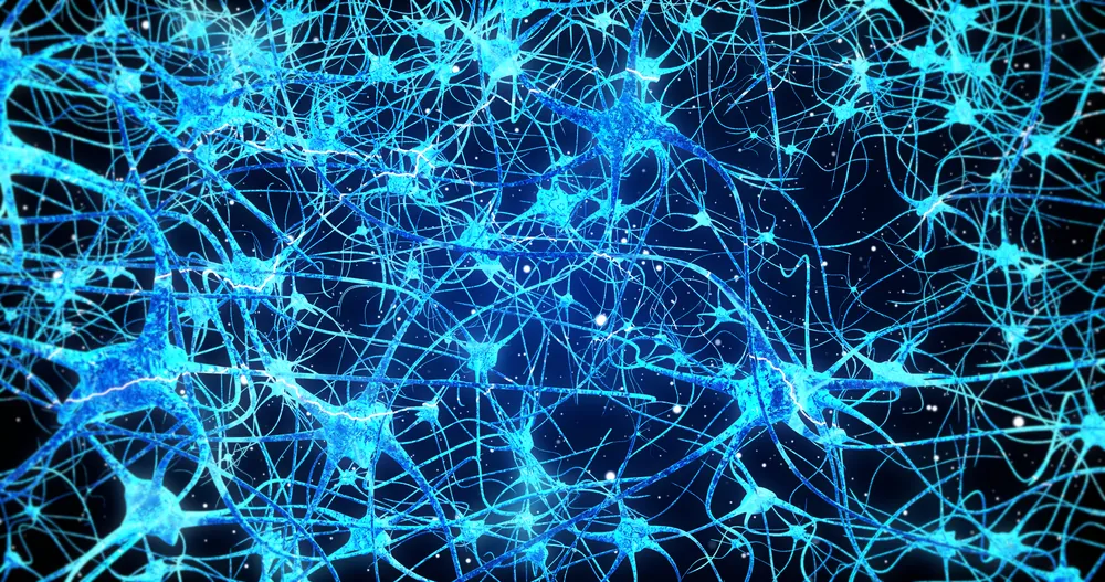
Our brain is mapping the world. Often that map is distorted, but it’s a map with constant immediate sensory input.
– EO Wilson
How do I use the Brain Map?
Therapy and psychometric testing is an imperfect attempt to see inside the brain from the outside. qEEG brainmapping can allow you to see inside the brain with less uncertainty, subjective error and clinical bias. The brain map can be used to create a neurostim plan but it can be used to do many other things to. Your brain map helps you and your therapist understand the way you think. It can provide objective proof of suspicions that you have about how your brain works and what it needs to heal.
The brain map can help your therapist understand what is happening in your brain and what treatment is best. Your thereapist is welcome to join us for the presentation of your brain map. You can do this virtually or in person. You can even do therapy with your therapist during neurostim, even if they are not at Taproot, while you recieve neurostimulation to reinforce the brain training.
Your brain map can help you understand yourself and your life in a different way. It can help you love and accept parts of you that you did not not understand and point you on the path to growth and healing. You can use it in therapy or individually to finally find what you are missing to grow and heal.
What are the Benefits of Neurostimulation?
Neurostimulation treats many psychological disorders and symptoms, but it can also have many secondary benefits. Neurostimulation can enhance creativity, reduce anxiety and depression, help academic performance, enhance focus and concentration, and help athletes maintain optimal athletic performance. Additionally, neurostimulation and neurofeedback can reduce the symptoms of bipolar disorder, manic depressive, OCD and ADHD permanently and without medication.
Is neurostimulation evidence based? Is there research about neurofeedback?
Yes! Even though the neurostimulation technology is state of the art and a cutting edge treatment for PTSD it has been used and researched for yeas by large institutions like The VA Veterans Administration, John’s Hopkin’s and The Mayo Clinic. Neurostimulation is non invasive and does not require surgery or have the side effects of medication. The results are often permanent.
You can read the most recent research on neurostimulation here.
What does Neurostimulation feel like?
Some people find neurostimulation relaxing, but most feel nothing durring neurostimulation sessions and notice the benefits after the session when symptoms reduce. Neurostimulation is a integrative and natural way to harness the brain’s own healing power. Neurostimulation harnesses the brains natural frequency responses to build new neural connections and reinforce positive neural pathways. Research shows that patients with TBI (traumatic brain injury) or memory loss have found that it can help the brain reroute connections around damage and heal damaged parts of the neural network.
Is neurostimulation and neurofeedback safe?
Yes! The FDA approved neurostimulation safe for consumer use and multiple research studies have confirmed it’s efficacy for a number of conditions. The technology is state of the art and new to the private sector but large institutions like Mayo Clinic, The VA, and John’s Hopkin’s have been using it for years. Now it is availible to you through collaboration between Peak Neuroscience and Taproot Therapy Collective.
Unlike other methods neurostimulation is naturaland holistic. It mimics the brains innate processes to harness the brain’s own natural healing ability. Neurostimulation recreates the processes of the growing and the developing brain so that the brain can learn new things like when you were a child!
Because Dr. Jay Mishalanie was such an early adopter of this new technology, he has more experience than many using this rare and exciting technology. Interpreting the brain maps and creating the stim plans is half art half science. This technology takes years to train in and Dr. Mishalaine was a pioneer in the neuroscience field with years of experience. You are in good hands!
How much does Neurostimulation cost?
Because there are so few people trained in reading the qEEG Brainmaps the price can vary based on availible clinical psychologists. The average cost is around $100 per session, however we run promos and discounts all the time! Sign up for our mailing list below to be alerted about specials that make therapy more affordable.
What Kind of Brain Waves Can QEEG Detect?
qEEG brain mapping is a powerful tool used by healthcare professionals to analyze various types of brain waves such as delta, alpha, theta, beta, and high beta waves. These waves, with their unique frequencies, provide valuable insights into a person’s neurological functioning and potential cognitive or mental health issues.
Delta Waves:
Delta waves are the slowest brain waves, with a frequency of 0.5-4 Hz. They are typically associated with deep sleep and can also be present in coma patients. The sensation of delta waves is often described as a profound state of relaxation, where the mind is in a state of rest and rejuvenation.
Theta Waves:
Theta waves have a frequency of 4-8 Hz and are typically observed during light sleep or drowsiness. They may also be present during meditation or creative activities. In qEEG brain mapping, an increase in theta waves may be associated with attention deficit hyperactivity disorder (ADHD), while a decrease in theta waves may be associated with cognitive decline in older adults. The sensation of theta waves is often described as a dreamy, introspective state.
Alpha Waves:
Alpha waves have a frequency of 8-12 Hz and are usually observed when a person is awake but relaxed or neutral. They are commonly experienced when closing the eyes or practicing meditation. Decreased alpha waves may be linked to anxiety or depression, while increased alpha waves may indicate improved relaxation and stress reduction. The sensation of alpha waves is often described as a state of calm and peacefulness.
Beta Waves:
Beta waves have a frequency of 12-30 Hz and are usually present when a person is awake and engaged in cognitive or physical activities. They are associated with alertness, focus, decision making, and concentration. Abnormalities in beta waves can be linked to conditions such as anxiety, depression, and insomnia. The sensation of beta waves is often described as a state of heightened awareness and mental activity.
High Beta Waves:
High beta waves have a frequency of 30-40 Hz and are often associated with intense cognitive or physical activities, such as problem-solving or exercise. An increase in high beta waves in qEEG brain mapping may be associated with conditions such as ADHD or obsessive-compulsive disorder (OCD). The sensation of high beta waves is often described as a state of heightened mental alertness and intense focus.
Summary of How qEEG Uses Brain Waves
The analysis of delta, alpha, theta, beta, and high beta waves in qEEG brain mapping can provide valuable information about a person’s neurological functioning and potential cognitive or mental health issues. The nuance in the brain map is the way we use these types of thinking and the interplay between them. By identifying abnormalities in these brain waves, healthcare professionals can develop more targeted and effective treatment plans for their patients. By understanding the uniqueness in each individual’s brain, healthcare professionals can develop targeted treatment plans for overall improved brain health.
The Relationship Between Glia and Mental Health
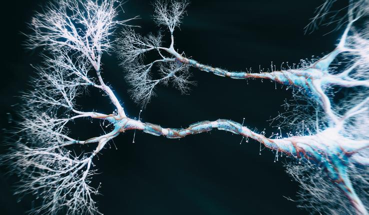
Microglia, often referred to as the immune cells of the brain, play a crucial role in maintaining mental health and regulating inflammation within the central nervous system. These specialized cells are responsible for monitoring brain tissue for abnormalities, pathogens, and damaged cells. Dysregulation of microglial function has been implicated in various mental health disorders including depression, anxiety, and neurodegenerative diseases like Alzheimer’s and Parkinson’s. Emerging technologies like quantitative electroencephalography (qEEG) brain mapping and neurostimulation offer promising avenues for enhancing microglial and myelin function. qEEG brain mapping allows for the precise assessment of brain activity, helping identify abnormal patterns that may indicate underlying mental health issues. Neurostimulation techniques, such as transcranial magnetic stimulation (TMS) or transcranial direct current stimulation (tDCS), have shown potential in modulating microglial activity and promoting the repair of myelin, the protective sheath around nerve fibers. By harnessing these innovative tools, researchers and clinicians are exploring new approaches to bolstering microglial function and addressing the intricate relationship between inflammation and mental well-being.
QEEG Brain Mapping and Neuromodulation: Optimizing the Brain’s Potential
QEEG (Quantitative Electroencephalography) Brain Mapping and Neuromodulation are cutting-edge approaches that allow us to understand and optimize brain function in a targeted, data-driven way. By measuring and analyzing brain wave patterns, we can identify areas of imbalance or inefficiency and then use neuromodulation techniques to gently guide the brain towards healthier, more integrated functioning.
The Science of QEEG Brain Mapping
QEEG Brain Mapping is a non-invasive process that uses EEG (electroencephalography) to measure the electrical activity of the brain. By placing sensors on the scalp, we can detect and record the brain’s rhythms and patterns with temporal and spatial precision.
The raw EEG data is then processed and analyzed using specialized software and normative databases. This allows us to create detailed “brain maps” that show how an individual’s brain activity compares to healthy norms for their age group. We can identify areas of over- or under-activation, asymmetries between brain regions, and patterns associated with various cognitive, emotional, and behavioral challenges.
This detailed snapshot of brain function provides invaluable insights for guiding therapy. As neuropsychologist Antonio Damasio has shown, our mental processes are inextricably linked to the physical substrates of the brain. By understanding the brain, we can more effectively understand and help the mind.
Neuromodulation: Training the Brain
With the insights of QEEG Brain Mapping, we can then use neuromodulation techniques like neurofeedback to gently guide the brain towards more optimal functioning. Neurofeedback is a form of biofeedback that uses real-time displays of brain activity to teach self-regulation.
During neurofeedback sessions, individuals are shown their own brain wave patterns and guided to modify them using mindfulness, visualization, and other techniques. With repeated practice, the brain can learn to operate in healthier, more efficient and flexible ways. This neuroplasticity-based training has been compared to physical therapy for the brain.
Other neuromodulation approaches, such as transcranial magnetic stimulation (TMS) and transcranial direct current stimulation (tDCS), use gentle magnetic or electrical currents to stimulate specific brain regions. This can help encourage underactive areas to come online or overactive areas to settle down into a healthier balance.
QEEG and Neuromodulation in Context
While QEEG and neuromodulation are grounded in modern neuroscience, they also connect with insights from psychology, anthropology, philosophy, and spirituality across cultures and eras.
The idea that we can measure and modify the physical substrates of the mind resonates with the ancient Greek maxim “know thyself” and the Buddhist concept of “mindfulness” – the ability to observe and shape one’s own mental processes. The focussed, meditative state cultivated in neurofeedback has parallels with practices from yoga to Christian mysticism.
Carl Jung and his followers used dream analysis, active imagination, and other techniques to map and engage with the deep psyche in ways that presage the biofeedback of neuromodulation. Jung’s concept of individuation – the drive towards wholeness and integration – also mirrors the goals of guiding the brain to more balanced, harmonious functioning.
In mythology and literature worldwide, there are resonant themes of descending into the underworld of the unconscious mind to retrieve hidden wisdom and healing. Dante’s journey through the afterlife in the Divine Comedy and the grail quests of Arthurian legend reflect a perennial human drive to illuminate the deep structures of the self – a drive QEEG and neuromodulation serve with modern tools.
Applications Across the Lifespan
By providing a window into brain function and a set of tools for optimization, QEEG Brain Mapping and neuromodulation can help with a wide range of goals and challenges across the lifespan:
- For those with ADHD, it can help improve focus, impulse control, and self-regulation
- For autism spectrum disorders, it can foster social engagement and communication
- For academic challenges, it can optimize learning, memory, and test performance
- For anxiety and trauma disorders, it can help rebalance the nervous system and build resilience
- For mood disorders, it can improve affect regulation and encourage more positive patterns
- For addictions, it can reduce cravings and impulsivity and strengthen healthier coping mechanisms
- For sleep disorders, it can help the brain settle into natural, restorative rhythms
- For peak performance, it can help people operate at their neurological best, whether in athletics, business, or creative pursuits
- For healthy aging, it can help keep the brain youthful and sharp
Empowering Brain and Mind
QEEG Brain Mapping and neuromodulation offer new ways to understand and enhance the most complex and powerful organ in the known universe – the human brain. By integrating cutting-edge science with the wisdom of the humanities, these approaches empower you to optimize your unique neurological gifts and reach your fullest potential.
If you’d like to learn more or experience the benefits of QEEG brain mapping and neuromodulation for yourself, please reach out. Our team would be honored to support you on your journey of growth and transformation.
Is qEEG Brainmapping Evidence Based?
ADHD and qEEG:
Research indicates that qEEG, particularly the measurement of the theta/beta ratio, has been widely studied in ADHD populations. It can help identify biomarkers that differentiate ADHD individuals from those without the disorder. Elevated theta power and reduced alpha power are the most consistently identified markers for ADHD, which helps guide neurofeedback interventions targeting attention regulation. Studies suggest that qEEG can also monitor treatment progress in ADHD by assessing brainwave abnormalities before and after interventions
.
Autism and qEEG:
Studies using qEEG have also identified patterns in the brain associated with autism spectrum disorder (ASD), especially in children. One study from the Kufa Medical Journal explored how qEEG can detect increased power in certain frequency bands, such as beta activity, which correlates with autistic symptoms. Neurofeedback sessions targeting abnormal alpha or mu bands in autistic children showed promising results in improving attention and reducing autism-related symptoms
. qEEG can be particularly useful in early diagnosis, even before behavioral symptoms fully manifest, by identifying unique brainwave patterns in high-risk newborns
.
Behavioral Regulation in Neurodevelopmental Disorders:
The Drake Institute provides evidence of how brain mapping (qEEG) can reveal dysfunctional brain areas contributing to behavioral issues in conditions like ADHD and autism. By analyzing dysregulated brain networks, targeted neurofeedback protocols have been shown to improve brain coherence, emotional regulation, and attention, providing long-term benefits beyond what is typically achieved with medication alone
Other Studies on qEEG Brain Mapping
-
Multicenter Trial for ADHD:
- A study replicating findings from 2012 confirmed that qEEG-informed neurofeedback led to a 76% response rate in ADHD symptom reduction. It highlighted better clinical outcomes when qEEG was tailored to individuals’ EEG profiles, significantly reducing inattention and hyperactivity
.
- qEEG for Autism Spectrum Disorders:
- A review noted qEEG’s role in early detection and treatment, helping to reduce cognitive and behavioral symptoms by identifying abnormal EEG patterns. Improvements were observed in social interactions and attention
.
-
Pediatric ADHD Study:
- qEEG was used to monitor neurofeedback efficacy in children with ADHD, with notable improvement in executive function and attention span
.
-
Neurofeedback for ADHD and Dopamine Dysfunction:
- Studies involving dopamine regulation through neurofeedback, guided by qEEG, showed significant symptom improvement in ADHD patients, particularly in managing impulsivity and attention control
.
-
Neurofeedback and Cognitive Functions in Autism:
- Research suggests qEEG-guided neurofeedback helps regulate neural patterns in autistic children, improving cognitive function, attention, and behavioral regulation
.
- EEG Subtypes and Neurofeedback Response in ADHD:
- Different EEG subtypes were studied to enhance neurofeedback’s effectiveness, with tailored treatment leading to higher remission rates, especially in children with anxiety and comorbid conditions
.
-
Brain Mapping and qEEG for ADHD:
- Clinics like the Drake Institute utilize qEEG brain mapping to identify areas of dysregulation in ADHD and autism, guiding non-pharmacological interventions that significantly improve behavioral outcomes
.
-
Longitudinal ADHD Neurofeedback Study:
- A long-term study using qEEG for neurofeedback in ADHD showed sustained improvements in attention and behavior, making it a promising alternative to medication
.
-
Neurofeedback for Behavioral Issues in Children:
- A trial involving children with behavioral dysregulation (due to ADHD or trauma) showed qEEG-informed neurofeedback led to substantial improvements in behavior control and emotional regulation
.
-
Combined Neurofeedback and Medication Approaches:
- Research highlights the benefit of combining qEEG-guided neurofeedback with traditional ADHD medications, optimizing both approaches for enhanced treatment outcomes
Best practices when finding a qeeg provider?
Credentials: Ensure the provider is certified and trained in qEEG interpretation and neurofeedback.
Experience: Look for a provider with extensive experience, particularly in your condition (ADHD, autism, etc.).
Technology: Check if they use up-to-date qEEG equipment and software.
Personalized Treatment: Opt for providers who offer individualized protocols based on your brain map.
Comprehensive Assessment: Choose a provider who performs a thorough qEEG assessment before treatment.
Multi-disciplinary Approach: Look for integration with other therapeutic methods (cognitive-behavioral therapy, medication, etc.).
Data Transparency: Ensure you receive clear reports and explanations of qEEG results.
Outcome Monitoring: Check if progress is regularly monitored through follow-up qEEG assessments.
Evidence-based Practices: Ask if they rely on research-supported methods for qEEG and neurofeedback.
Tailored Neurofeedback: Verify that neurofeedback sessions are personalized based on qEEG data.
Consultation Availability: Make sure they offer initial consultations to discuss qEEG and treatment options.
Referrals and Reviews: Seek out referrals or reviews from previous patients.
Ethical Standards: Ensure they follow ethical guidelines for patient treatment and data privacy.
Insurance Coverage: Check whether they accept insurance or offer payment plans.
Long-term Support: Look for a provider that offers long-term treatment plans and support after qEEG sessions.
Why Choose NeuroField Technology
NeuroField technology offers a unique combination of qEEG-guided neurofeedback, neuromodulation, and pulsed electromagnetic field (PEMF) therapy to optimize brain function. It integrates cutting-edge tools to improve neurological health, making it especially effective for treating conditions like ADHD, autism, and anxiety. NeuroField’s multi-modal approach tailors treatments to individual brainwave patterns, improving efficacy and outcomes. This makes it an excellent option for those seeking personalized, research-backed interventions to enhance cognitive and emotional regulation.
NeuroField doesn’t just target symptoms—it addresses underlying brainwave dysfunctions by recalibrating abnormal brainwave activity. Neurofeedback enhances the brain’s natural ability to regulate itself, leading to longer-lasting improvements in cognitive and emotional functioning. By combining multiple technologies, including PEMF to stimulate the body’s natural healing processes, NeuroField provides a holistic treatment that can improve energy levels, mood stability, and attention span.
One of the main benefits of NeuroField is its non-invasive nature, making it an excellent option for those seeking alternatives to medication. It is supported by research that shows its efficacy in reducing symptoms of neurodevelopmental disorders, trauma, and stress-related conditions. Additionally, NeuroField technology is highly adaptable, allowing for continuous adjustments based on real-time feedback from the brain, ensuring optimal treatment progression. For individuals seeking a scientifically grounded and customized therapy, NeuroField offers a promising pathway toward enhanced neurological health.
What are the Parts of the qEEG Brain Map?
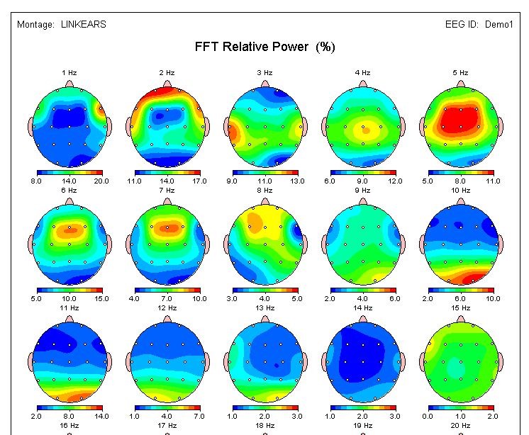
The qEEG brain map results provide information about different brain speeds, such as delta, theta, alpha, beta, and high beta, which correspond to different states based on circadian rhythms. Colors on the map indicate whether the brain is using these speeds at higher or lower levels than optimal. The top row of heads on the map represents the overall power of each speed, while the relative power shows which speed is being used the most and the least in comparison to other maps of people that match your age and gender assigned at birth.
The parameters at the bottom of the map, including amplitude, asymmetry, coherence, and phase lag, represent the communication between different brain areas, similar to networks in the brain. The dots on the map represent different areas of the brain:
- F for frontal areas responsible for attention and executive function
- C for central areas responsible for basic functionality and the body’s nervous systems
- T for temporal areas responsible for auditory processing and emotional regulation
- O for occipital areas responsible for visual processing
A close-to-optimal map with minimal lines indicates efficient communication between brain areas in this example. Overall, qEEG provides valuable information about the functioning of the human brain and can help in understanding brain patterns and states.
What is an Example of a qEEG Brain Map?
qEEG brain maps can provide valuable information about the electrical activity of the brain and can help identify patterns or abnormalities that may be associated with various neurological or psychiatric conditions. Here are some examples of what a qEEG brain map might show:
Example of a qEEG Brain Map: ADHD
Let’s say a patient presents with symptoms of attention deficit hyperactivity disorder (ADHD), such as difficulty concentrating, impulsivity, and hyperactivity. While each person’s brain will show different results, the qEEG brain map may reveal an excess of theta (4-8 Hz) waves and a deficiency of beta (12-30 Hz) waves in the prefrontal cortex. This is the region of the brain responsible for executive functions such as attention, decision-making, and impulse control.
This pattern of excess theta and deficient beta activity in the prefrontal cortex has been associated with ADHD in previous research. Based on this qEEG finding, a healthcare professional may recommend neurostimulation and neurofeedback therapy to help train the patient’s brain to increase beta activity in the prefrontal cortex and decrease theta activity; potentially leading to a reduction in ADHD symptoms.
It’s important to note that qEEG brain maps should be interpreted by a qualified healthcare professional who has expertise in qEEG, neurostimulation, and neurofeedback. The interpretation of qEEG results should take into account the patient’s clinical history and other factors, and the results should not be used to make a diagnosis without additional evaluation.
Example of a qEEG Brain Map: Autism
An analysis of a child’s brain using a qEEG brain map might reveal an atypical pattern of electrical activity in the temporal and frontal lobes, which are regions crucial for social queues, communication, and language processing. Based on these qEEG results, a healthcare professional may suggest neurostimulation and neurofeedback therapy as a potential intervention. The goal of this therapy would be to train the child’s brain to reduce theta and alpha activity in the temporal and frontal lobes, which could potentially enhance their language processing, processing social queues, and communication abilities. The therapy might involve engaging the child in interactive games or exercises that offer real-time neurofeedback on their brain activity, aiding them in learning self-regulation of their brain function.
Example of a qEEG Brain Map: Chronic Pain
Imagine a patient grappling with chronic pain that persists despite conventional treatments like medications and physical therapy. An innovative approach utilizing a qEEG brain map might shed light on the issue by uncovering an intriguing pattern of abnormal electrical activity within the somatosensory cortex. This particular region of the brain plays a vital role in processing sensory information; encompassing touch, pressure, and pain.
Notably, the qEEG assessment may unveil heightened activity in the delta (0-4 Hz) and alpha (8-12 Hz) frequency bands within the somatosensory cortex. Fascinatingly, these frequency bands have been closely associated with pain processing and the complex experience of chronic pain.
Drawing from this remarkable qEEG discovery, a healthcare professional may introduce the concept of neurostimulation and neurofeedback therapy, an empowering avenue to train the patient’s brain to reduce delta and alpha activity within the somatosensory cortex. This can potentially lead to a remarkable alleviation of their pain perception. Neurostimulation and neurofeedback therapies, an embodiment of technological ingenuity, enable the patient to receive real-time feedback regarding their brain’s activity; effectively empowering them to master the art of self-regulating their brain function. This captivating therapy holds the promise of transforming the patient’s relationship with pain and bestowing them with newfound control over their mental and physical well-being.
What is an Example of a Neurostimulation Plan?
NeuroField is a type of neurostimulation that uses low-intensity electromagnetic fields to modulate brain activity. The device can be programmed to target specific regions of the brain and modulate the activity of neurons in those regions. Provided below are two examples of Neurofield Neurostimulation plans and what would be modulated.
Example of a Neurostimulation plan for Parkinson’s Disease:
Target region for parkinson’s disease:
The primary motor cortex (PMC) is a region of the brain that is involved in the planning and execution of voluntary movements. Abnormal activity in the PMC has been implicated in various movement disorders, such as Parkinson’s disease.
Modulation for parkinson’s disease:
The NeuroField device would be programmed to deliver low-intensity electromagnetic fields to the PMC to modulate its activity. The fields would be timed and calibrated to match the natural rhythm of the individual’s brain activity in the PMC. This would help to normalize the activity of neurons in the PMC and improve movement control.
Treatment sessions for parkinson’s disease:
The patient would attend multiple treatment sessions, each lasting approximately 20-30 minutes. During the session, the NeuroField device would be placed on specific regions of the head, using a cap, to target the PMC. The patient may feel a mild sensation of warmth or tingling during the treatment, but it should not be painful or uncomfortable.
Follow-up evaluations for parkinson’s disease:
After the treatment sessions are complete, the patient would undergo a follow-up evaluation to assess changes in symptoms and to determine if additional sessions are needed. A qEEG brain map may be repeated to evaluate changes in brain activity and to guide further treatment.
Example of a Neurostimulation plan for Major Depressive Disorder:
Target region for major depressive disorder:
The dorsolateral prefrontal cortex (DLPFC) is a region of the brain that is involved in cognitive control and emotional regulation. Abnormal activity in the DLPFC has been implicated in major depressive disorder.
Modulation for major depressive disorder:
The NeuroField device would be programmed to deliver low-intensity electromagnetic fields to the DLPFC to modulate its activity. The fields would be timed and calibrated to match the natural rhythm of the brain’s activity in the DLPFC. This would help to normalize the activity of neurons in the DLPFC and improve emotional regulation.
Treatment sessions for major depressive disorder:
The patient would attend multiple treatment sessions, each lasting approximately 20-30 minutes. During the session, the NeuroField device would be placed on specific regions of the head to target the DLPFC. The patient may feel a mild sensation of warmth or tingling during the treatment, but it should not be painful or uncomfortable.
Follow-up evaluations for major depressive disorder:
After the treatment sessions are complete, the patient would undergo a follow-up evaluation to assess changes in symptoms and to determine if additional sessions are needed. A qEEG brain map may be repeated to evaluate changes in brain activity and to guide further treatment.
Example of a Neurostimulation plan for PTSD and Anxiety:
Target region for PTSD and Anxiety:
The amygdala is a region of the brain that plays a key role in the processing of emotions; particularly fear and anxiety. Abnormal activity in the amygdala has been implicated in anxiety disorders.
Modulation for PTSD and Anxiety:
The NeuroField device would be programmed to deliver low-intensity electromagnetic fields to the amygdala to modulate its activity. The fields would be timed and calibrated to match the natural rhythm of the brain’s activity in the amygdala. This would help to normalize the activity of neurons in the amygdala and work to reduce anxiety.
Treatment sessions for PTSD and Anxiety :
The patient would attend multiple treatment sessions, each lasting approximately 20-30 minutes. During the session, the NeuroField device would be placed on specific regions of the head to target the amygdala. The patient may feel a mild sensation of warmth or tingling during the treatment, but it should not be painful or uncomfortable.
Follow-up evaluations for PTSD and Anxiety:
After the treatment sessions are complete, the patient would undergo a follow-up evaluation to assess changes in symptoms and to determine if additional sessions are needed. A qEEG brain map may be repeated to evaluate changes in brain activity and to guide further treatment.
Additional Information on Neurostimulation Plans:
It’s important to note that the use of NeuroField for anxiety disorders is still being studied, and more research is needed to determine its effectiveness and safety. The neurostimulation plan should be conducted under the guidance of a qualified healthcare professional who has expertise in qEEG and neurostimulation. The treatment plan should be tailored to the individual patient’s needs and should take into account the patient’s clinical history and other factors.
How is the qEEG Brain Map Analyzed?
By capturing functional images of the brain’s electrical waves, qEEG brain maps offer valuable information about brain patterns and states. Many people are curious how the brain maps are interpreted. The process of interpreting and analyzing qEEG brain maps takes years to learn and the technology is so new that few people have been trained in reading them. Interpreting the maps is half art half science. Dr. Jason Mishalanie, PhD, BCN was an early adopter of the technology and has more experience than almost anyone in the field.
Interpretation of qEEG Brain Maps:
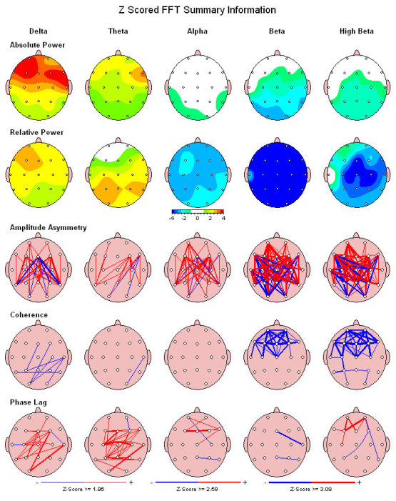
qEEG brain maps are generated by analyzing the electrical activity of the brain recorded through specialized caps with multiple electrodes placed on the scalp using a special cap. These maps typically display different brain speeds and waves, including delta, theta, alpha, beta, and high beta, which correspond to different states based on circadian rhythms. Interpretation of these brain speeds and waves involves analyzing the colors displayed on the map, which indicate whether the brain is using these speeds at higher or lower levels than optimal.
Colors on the qEEG brain map:
The colors on the qEEG brain map play a crucial role in interpreting the brain’s activity. Yellow, orange, and red colors indicate that the brain is using one to three levels too high of a particular speed, while different blue colors suggest that the brain is using one to three levels too low of that speed. This color-coded information helps in identifying any imbalances or irregularities in brain activity, providing valuable insights into the functioning of the brain.
Overall power and relative power:
The top row of heads on the qEEG brain map represents the overall power of each brain speed, indicating how ‘charged up’ the brain is overall. This information helps in understanding the overall activity levels of different brain speeds. Additionally, the relative power displayed on the map shows which brain speed is being used the most and the least in comparison to others of the same age and gender assigned at birth. This data provides important clues about the brain’s dominant and less dominant activity levels, aiding in the interpretation of qEEG brain maps.
Parameters at the bottom of the map:
The qEEG brain maps also include parameters at the bottom of the map that provide insights into the communication between different brain areas. These parameters, including amplitude, asymmetry, coherence, and phase lag, represent the networks in the brain and how different areas communicate with each other. For instance, frontal areas responsible for attention and executive function are labeled with “F,” central areas with “C,” temporal areas with “T,” and occipital areas with “O.” The analysis of these parameters and the lines connecting different areas on the map help in understanding the efficiency of communication between brain regions.
Z-Score:
The Z-score coherence is a measure of functional connectivity between two regions of the brain. It provides an estimate of the strength of the coherence between the signals recorded from different electrode sites, compared to a database. The coherence is a measure of the degree to which two signals are synchronized or correlated, indicating the degree of functional connectivity between different brain regions. The Z-score is a statistical measure of how far the coherence value is from the average coherence value in the normative database.
The Z-score amplitude is a measure of the power or strength of the electrical activity in a particular frequency band within a specific region of the brain. The amplitude is the measurement of the size or magnitude of a particular qEEG wave. The Z-score amplitude is the statistical comparison of the amplitude value of a particular frequency band within a specific region of the brain compared to a normative database.
Both Z-score coherence and amplitude are useful in the assessment of brain function and dysfunction. They can provide valuable information about the patterns of brain activity associated with various neurological and psychiatric conditions, such as attention deficit hyperactivity disorder (ADHD), depression, anxiety, and traumatic brain injury. Z-score coherence and amplitude can also be used to guide neurostimulation treatments to target specific brain regions and frequencies for optimal outcomes.
Amplitude Asymmetry:
Amplitude asymmetry refers to the difference in the electrical activity between the left and right hemispheres of the brain. It is typically measured as the difference in amplitude between homologous electrode sites located on each hemisphere. An abnormal amplitude asymmetry may suggest a disruption in the normal functioning of the brain, and has been associated with various neurological and psychiatric conditions, including depression, anxiety, and schizophrenia.
Phase Lag:
Phase lag is a measure of the delay in the propagation of neural signals between different regions of the brain. It is a measure of the temporal relationship between two or more qEEG signals recorded from different electrode sites. Phase lag is typically calculated by measuring the time delay between two signals at a given frequency. An abnormal phase lag may suggest a disruption in the normal communication between different brain regions, and has been associated with various neurological and psychiatric conditions, including attention deficit hyperactivity disorder (ADHD), autism, and traumatic brain injury.
Implications of qEEG Brain Map Interpretation:
Interpretation of qEEG brain maps can have significant implications for understanding brain function and identifying any abnormalities or imbalances in your brain. By analyzing the brain’s activity levels, dominant and less dominant patterns, and communication between the different brain areas, qEEG brain maps can provide valuable insights into the functioning of the human brain. This information can be used in various clinical and research settings, such as identifying neurological disorders, monitoring treatment progress, and optimizing cognitive performance.
Summary of how the QEEG map is analyzed:
qEEG brain maps are a powerful tool for interpreting and analyzing the functional activity of the brain. By analyzing the colors on the map, overall power and relative power of brain speeds, and parameters related to communication between brain areas, qEEG brain maps can provide valuable insights into brain function. Understanding the interpretation of qEEG brain maps can help in optimizing brain health, identifying neurological disorders, and improving cognitive and athletic performance.
The Myers-Briggs Type Indicator (MBTI) and qEEG Brain Mapping
The Myers-Briggs Type Indicator (MBTI) is a widely used personality assessment that categorizes individuals into 16 distinct personality types based on four dichotomies: extraversion vs. introversion, sensing vs. intuition, thinking vs. feeling, and judging vs. perceiving. Quantitative Electroencephalography (qEEG) brain mapping is a diagnostic tool used to measure and map brainwave activity across different regions of the brain. Researchers have explored potential connections between these two domains to establish a relationship between them.
Several researchers have proposed that the various brainwave frequencies observed in a qEEG brain map may correspond to the functions identified in the MBTI. However, the precise relationship between qEEG brain waves and MBTI functions remains a subject of research and debate.
One proposed connection suggests that the alpha brainwave frequency, associated with relaxed wakefulness and meditation, is linked to the MBTI function of intuition. Alpha waves reflect a state of relaxed focus that fosters insight and creativity, which may facilitate the intuition function involving generating insights and making connections based on patterns and associations.
Another proposed connection suggests that the beta frequency, associated with focused attention and alertness, may correspond to the MBTI function of sensing. Beta waves reflect a state of focused attention that enables precise and detailed perception, potentially facilitating the sensing function of gathering data through the senses and paying attention to concrete details and facts.
Furthermore, the theta frequency, associated with daydreaming and creative states, is purported to correspond to the MBTI function of feeling. Theta waves reflect a state of relaxed and open awareness, fostering creative and imaginative thinking that may facilitate the feeling function of evaluating and assessing information based on personal values and emotional responses.
Likewise, the delta frequency, associated with deep sleep and unconscious processing, may correspond to the MBTI function of thinking. Delta waves reflect a state of unconscious processing that supports problem-solving and decision-making, potentially facilitating the thinking function of analyzing and evaluating information based on logic and reason.
However, it is important to note that while some correlations between qEEG brain waves and MBTI functions have been proposed, conclusive evidence for these connections is lacking. The brain is a complex and dynamic system, and it is unlikely that a single brainwave frequency can fully account for a specific cognitive or personality function. Additionally, the MBTI relies on self-report assessments, introducing biases and limitations.
Nonetheless, exploring the potential connections between the different brain waves observed in a qEEG brain map and the functions identified in the MBTI can yield valuable insights into the relationship between brain activity and personality.
Trauma and its Impact on Diagnosis, Symptoms, and Personality
Trauma is a widespread phenomenon that can have long-lasting effects on an individual’s psychological and physical well-being. While genetics plays a significant role in shaping personality traits and behavior, research indicates that environmental factors, including trauma, can also influence their expression. Recent studies have focused on understanding the intricate interplay between genetic predisposition and environmental factors, specifically how trauma can lead to changes in genetic expression.
Trauma’s influence on genetic expression is primarily mediated through epigenetic mechanisms. Epigenetic changes involve alterations in gene expression without modifications to the DNA sequence itself, but rather changes in gene regulation. Trauma can induce epigenetic modifications that alter gene expression, resulting in the manifestation of symptoms associated with mental health conditions.
The Dresden study, conducted by scientists at the Max Planck Institute of Psychiatry in Germany, examined the epigenetic changes occurring in individuals with Post-Traumatic Stress Disorder (PTSD). The study revealed that PTSD can trigger epigenetic modifications in genes associated with stress response, immune function, and neuronal signaling pathways. These changes can lead to alterations in brain function and structure, contributing to the development of PTSD symptoms.
Additionally, research has demonstrated that early childhood trauma can induce epigenetic changes in genes responsible for regulating the stress response system, increasing the risk of developing anxiety and depression. Similarly, trauma can impact the expression of genes related to addiction, rendering individuals more vulnerable to substance abuse and dependence.
Furthermore, trauma can result in changes in brain function and structure, leading to modified neural pathways underlying the expression of mental health symptoms. Studies have indicated that exposure to trauma can bring about alterations in brain regions responsible for emotional processing, consequently giving rise to anxiety and mood disorders.
Another study carried out by researchers at the University of Michigan observed that individuals with high scores in the trait of “neuroticism” were more prone to experiencing traumatic events and developing PTSD symptoms compared to those with low scores in neuroticism. Neuroticism is a personality trait characterized by a tendency to experience negative emotions such as anxiety, depression, and self-doubt.
In addition to personality, kindness, and compassion can also influence the experience and manifestation of trauma-related symptoms. Research has shown that individuals who engage in acts of kindness and compassion towards themselves and others may be better equipped to cope with the effects of trauma. For instance, a study conducted at the University of Arizona demonstrated that individuals who practiced self-compassion were less likely to experience PTSD symptoms following a traumatic event. Similarly, another study found that individuals who engaged in acts of kindness towards others experienced lower levels of stress and anxiety.
These findings underscore the significance of considering the role of personality and positive psychological factors such as kindness and compassion in the experience and expression of trauma-related symptoms. While genetics may determine how an individual responds to trauma and subsequent mental health conditions, environmental factors such as personality traits and positive psychological factors also play a critical role in shaping the manifestation of these symptoms.
We have multiple clinicians availible at Taproot Therapy Collective that treat a wide variety of issues and conditions with training in many techniques and modalities of therapy.
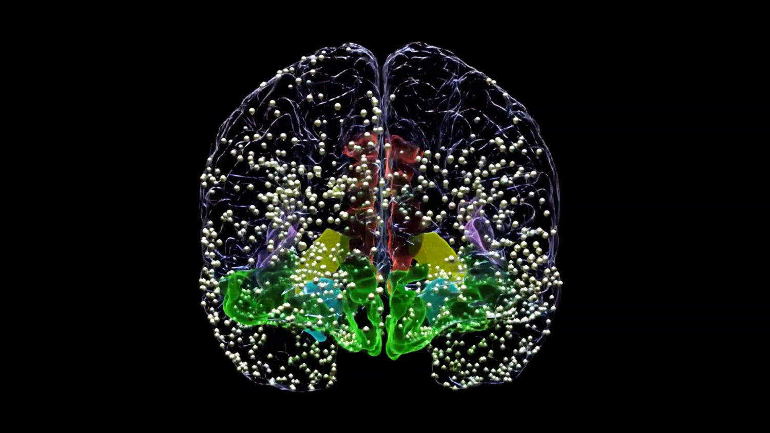
The History of Neurostimulation
Ancient World:
The history of neurostimulation spans ancient times to the present, with significant developments and pioneers driving its progress. Ancient civilizations, like the Greeks, experimented with electric sea creatures for treating ailments. In the Enlightenment era, medical electricity gained popularity, and researchers like Johann Gottlob Krüger and Matthias Bose explored its potential therapeutic applications.
Early History:
In the twentieth century, neurostimulation took significant strides. Dr. Albert Grass developed the first neurostimulator in the 1920s, leading to its use in treating epilepsy and chronic pain. Dr. G. W. Crile introduced electrical currents for chronic pain management, and Dr. Hans Selye explored the physiological responses of the nervous system to stress using electrical stimulation.
The 1950s saw the introduction of implanted neurostimulators, with Dr. William Sweet using implanted electrodes for chronic pain treatment. In 1967, the first spinal cord stimulator was developed, offering relief for chronic pain sufferers. The 1980s brought deep brain stimulation (DBS) for Parkinson’s disease, revolutionizing treatment options and improving patients’ quality of life.
Modern Advancements:
Modern advancements include non-invasive alternatives like transcranial magnetic stimulation (TMS), which uses magnetic fields to treat depression. The early 2000s saw the introduction of non-invasive spinal cord stimulators, offering targeted pain relief without surgery. Closed-loop systems and real-time adjustments based on brain activity monitoring have opened doors for personalized and adaptive neurostimulation.
Pioneers in the Field of Neurostimulation:
Today, neurostimulation is widely used to treat various conditions, including chronic pain, epilepsy, depression, and obsessive-compulsive disorder. Notable pioneers in the field include Dr. Benjamin Franklin, Dr. Melvin D. Yahr, Dr. Alim-Louis Benabid, and Dr. Mark S. George. Their contributions have paved the way for advancements in neurostimulation technology, with ongoing research promising further breakthroughs and improved treatments for neurological disorders. Neurostimulation continues to hold great potential for enhancing the lives of patients and shaping the future of medical care.
Contemporary qEEG Researchers:
Dr. Barry Sherman has been involved in groundbreaking research to enhance the understanding and diagnosis of mental health disorders. His research efforts have focused on utilizing neuroimaging techniques, such as functional magnetic resonance imaging (fMRI) and electroencephalography (EEG), to uncover the neurobiological underpinnings of various conditions. By studying brain activity and connectivity, he has contributed to the identification of distinct biomarkers and neurophysiological abnormalities associated with mental health disorders, including depression, anxiety, schizophrenia, and attention-deficit/hyperactivity disorder (ADHD).
Dr. Robert Thatcher: Dr. Thatcher is a leading expert in qEEG and has conducted extensive research on brainwave patterns and their relationship to mental health disorders. His work focuses on qEEG-guided neurofeedback, brain mapping, and the neurophysiology of various conditions, including ADHD, autism spectrum disorders, and traumatic brain injury.
Dr. Juri Kropotov: Dr. Kropotov has made significant contributions to the field of qEEG in mental health, particularly in the areas of depression, schizophrenia, and ADHD. His research explores the use of qEEG phenotypes for diagnosis, treatment planning, and monitoring treatment outcomes.
Dr. Martijn Arns: Dr. Arns has conducted extensive research on qEEG and its applications in mental health disorders. His work focuses on neurofeedback interventions for conditions such as ADHD, depression, and anxiety disorders. He has also contributed to the development of personalized treatment protocols based on qEEG assessments.
Dr. Juri D. Kropotov: Dr. Kropotov’s research focuses on the use of qEEG in the assessment and treatment of various mental health conditions, including attention disorders, mood disorders, and anxiety disorders. His work involves the development of novel qEEG-based interventions and the investigation of neurophysiological markers in psychiatric disorders.
Dr. Marco Congedo: Dr. Congedo has made significant contributions to the field of qEEG analysis and its applications in mental health. His research focuses on the development of advanced qEEG methods, machine learning algorithms, and the use of qEEG in the diagnosis and treatment of psychiatric disorders, including depression and anxiety.
Contemporary qEEG Applications:
qEEG Phenotypes:
qEEG phenotypes refer to patterns and characteristics of brainwave activity that are identified through the analysis of Quantitative Electroencephalography (qEEG) data. qEEG is a neuroimaging technique that measures and records the electrical activity of the brain using electrodes placed on the scalp. By analyzing the resulting data, researchers and clinicians can identify specific brainwave patterns that are associated with different mental states, cognitive processes, and mental health disorders.
qEEG phenotypes provide valuable insights into the functioning of the brain and its relationship to various aspects of mental health. They offer a quantitative and objective way to assess brain activity, allowing for a deeper understanding of brain function and dysfunction.
These phenotypes can be categorized based on the frequencies and amplitudes of brainwave activity recorded by qEEG. Common brainwave frequencies include delta (0.5-4 Hz), theta (4-8 Hz), alpha (8-12 Hz), beta (12-30 Hz), and gamma (30-100 Hz). Variations in these frequencies and their distribution across different brain regions can provide valuable information about an individual’s cognitive processes, emotional states, and potential abnormalities.
qEEG phenotypes have numerous applications in mental health. They can assist in the diagnosis of various mental health disorders, including depression, anxiety, attention-deficit hyperactivity disorder (ADHD), and post-traumatic stress disorder (PTSD). By comparing an individual’s qEEG data to normative databases or established patterns, clinicians can identify deviations and abnormalities that may indicate specific conditions.
qEEG phenotypes play a crucial role in personalized treatment planning. They can guide the selection of appropriate therapies, such as neurofeedback training, where individuals learn to self-regulate their brainwave activity based on the feedback provided by the qEEG. qEEG phenotypes can also aid in optimizing medication management by identifying specific brainwave patterns associated with treatment response to different medications.
qEEG phenotypes provide objective and quantifiable information about brain activity and its relevance to mental health. By leveraging these insights, researchers and clinicians can enhance diagnostic accuracy, tailor interventions, and improve outcomes in the field of mental health.
QEEG Phenotypes in Mental Health:
Diagnosis and Treatment Planning:
qEEG phenotypes enable clinicians to observe patterns and deviations in brainwave activity associated with various mental health disorders, such as depression, anxiety, attention-deficit/hyperactivity disorder (ADHD), and post-traumatic stress disorder (PTSD). This objective data aids in accurate diagnosis and personalized treatment planning.
Neurofeedback and Brain Training:
qEEG-guided neurofeedback therapy allows individuals to learn how to self-regulate their brainwave activity. By providing real-time feedback on their brainwave patterns, individuals can develop techniques to enhance desirable brainwave states, leading to improved mental health outcomes.
Medication Optimization:
qEEG phenotypes can assist psychiatrists in selecting the most appropriate medication for patients. By analyzing an individual’s qEEG data, clinicians can identify specific brainwave patterns that correlate with treatment response to particular medications, facilitating a more personalized and effective medication management approach.
Performance Enhancement:
qEEG phenotypes have applications beyond mental health disorders. Athletes, executives, and artists can benefit from qEEG assessments to optimize their cognitive and creative abilities. By identifying areas of brain activity that may need improvement, tailored interventions can be designed to enhance performance in specific domains.
Benefits of qEEG Phenotypes:
Objective and Quantifiable Data:
qEEG phenotypes provide objective and quantifiable data about brain activity, enhancing diagnostic accuracy and treatment efficacy. This data-driven approach allows for personalized and targeted interventions, leading to improved outcomes.
Early Intervention and Prevention:
qEEG phenotypes offer the potential for early identification of at-risk individuals before the onset of significant symptoms. This early intervention can promote proactive mental health care and prevent the exacerbation of conditions.
Non-Invasive and Safe:
qEEG is a non-invasive procedure that poses minimal risk to patients. The process involves attaching electrodes to the head using a special cap. The recording process has little to no feeling, making it suitable for individuals of all ages.
Holistic Approach to Mental Health:
qEEG phenotypes provide a comprehensive understanding of brain function and facilitate a holistic approach to mental health treatment. By considering the individual’s unique neurophysiology, interventions can be tailored to address underlying imbalances, fostering long term well-being.
As our understanding of the brain continues to evolve, qEEG phenotypes have emerged as a valuable tool in the field of mental health. By leveraging the power of neuroimaging and data analysis, mental health practitioners can enhance diagnosis accuracy, personalize treatment plans, and empower individuals on their journey toward optimal mental well-being. Embracing the potential of qEEG phenotypes, we can unlock a new era of targeted and effective mental health care.
Our Other Therapy Methods
QEEG Brain Mapping, Neurofeedback, and Neurostimulation: A Comprehensive Guide
At Taproot Therapy Collective in Hoover, Alabama, we offer advanced brain-based treatments including QEEG brain mapping, neurostimulation, and neurofeedback. These cutting-edge neurological technologies can help address a wide variety of mental health conditions by targeting the actual neurological sources of symptoms, rather than just managing the symptoms themselves.
Table of Contents
What is QEEG Brain Mapping?
Quantitative Electroencephalography (QEEG) brain mapping is a non-invasive diagnostic technology that measures electrical activity in the form of brain wave patterns. Unlike traditional brain imaging (such as MRI or CT scans) that show structural information, QEEG reveals how your brain is actually functioning in real-time.
During a QEEG brain mapping session at Taproot Therapy Collective, we place a comfortable cap with 19 sensors on your head to record brain activity. This process is entirely painless and takes approximately 30 minutes. The cap records electrical signals from different areas of your brain, measuring various brainwave frequencies (delta, theta, alpha, beta, and high beta) and how these brain regions communicate with each other.
The resulting data creates a detailed map of your brain's electrical activity that shows:
- Areas of optimal brain function
- Areas that may be overactive or underactive
- Communication patterns between different brain regions
- Imbalances that may be contributing to your symptoms
This brain map serves as a "fingerprint" of your unique brain patterns and provides critical information that helps us develop highly personalized treatment protocols using neurostimulation and neurofeedback. The QEEG brain map gives us objective data about what's actually happening in your brain, rather than relying solely on observed symptoms or subjective reports.
As Dr. Jason Mishalanie, PhD, BCN, our brain mapping specialist at Taproot Therapy Collective explains, "QEEG brain mapping provides a window into brain functioning that allows us to identify the source of symptoms or dysregulation. This helps us establish maximally effective neurofeedback treatment protocols for helping patients improve their quality of life without having to rely on medications."
Understanding Neurostimulation
Neurostimulation is an advanced therapeutic technique that uses targeted electrical or electromagnetic stimulation to modulate brain activity. At Taproot Therapy Collective, our neurostimulation approach is personalized based on your QEEG brain map results.
Unlike older technologies like transcranial magnetic stimulation (TMS) or microcurrent neurofeedback (MCNF) that use a "one-size-fits-all" approach to stimulate the entire brain with the same frequency, our neurostimulation techniques are precisely targeted. We can deliver different frequencies to different parts of the brain simultaneously, based on your specific needs.
During a neurostimulation session, you wear a comfortable cap with electrodes that can mimic the frequency of neurons in your brain. The cap delivers gentle electrical signals that "speak" directly to your neurons using frequencies called phases—which replicate the natural way neurons communicate with each other.
This targeted stimulation can:
- Open new neural pathways in the brain
- Teach the brain new ways of functioning
- Regulate overactive or underactive brain regions
- Improve communication between different brain areas
- Enhance focus, concentration, emotional regulation, and more
What sets our approach apart is that neurostimulation at Taproot Therapy is entirely personalized to your brain's unique patterns as identified in your QEEG brain map. This level of customization helps explain why many patients experience significant improvements even when other treatments, including medications, have failed to provide relief.
For more detailed information about what neurostimulation feels like, you can read our article: What Does QEEG Brain Mapping and Neurostimulation Feel Like?
The Neurofeedback Process
After neurostimulation opens new neural pathways in the brain, neurofeedback helps strengthen and reinforce these connections. Neurofeedback is a form of biofeedback specifically focused on brain activity, where you learn to self-regulate your brain function based on real-time feedback.
During a neurofeedback session at Taproot Therapy Collective, your brain activity is monitored in real-time while you receive feedback—typically through visual or auditory cues—when your brain produces desired patterns. Through repeated sessions, your brain learns to maintain healthier patterns, similar to how muscles develop through exercise.
This process can lead to potentially permanent improvements without medication or expensive long-term therapy. The brain essentially learns to function in more optimal ways, creating lasting neurological changes that can significantly reduce or eliminate symptoms.
Neurofeedback differs from neurostimulation in that:
- Neurostimulation actively introduces signals to the brain
- Neurofeedback trains the brain to regulate itself
- Together, they create a powerful approach to brain-based healing
Our specialized approach combines both technologies for maximum effectiveness, first using neurostimulation to open neural pathways and then using neurofeedback to strengthen and maintain these new, healthier patterns.
Conditions That Can Be Treated
QEEG brain mapping, neurostimulation, and neurofeedback have shown effectiveness for numerous conditions affecting children, adolescents, and adults. At Taproot Therapy Collective, we regularly help patients with:
Attention and Learning
- ADHD and ADD without medication
- Autism Spectrum Disorder (ASD)
- Learning disabilities
- Academic performance issues
- Executive functioning problems
Mood and Anxiety Disorders
- Depression
- Anxiety disorders
- Panic disorders
- Bipolar disorder
- Post-partum depression
Trauma and Stress
Physical and Performance
- Chronic pain
- Sleep disorders
- Athletic performance enhancement
- Cognitive enhancement
- Creativity enhancement
The applications of these technologies continue to expand as research advances. Patients often report improvements not only in their primary symptoms but also in overall mental clarity, emotional regulation, sleep quality, and general wellbeing.
Understanding Brain Waves
Your brain is constantly generating electrical activity in the form of brain waves. Different types of brain waves correspond to different mental states and cognitive functions. QEEG brain mapping measures these waves to identify patterns associated with various conditions.
| Brain Wave | Frequency | Associated With | When Imbalanced |
|---|---|---|---|
| Delta | 0.5-4 Hz | Deep sleep, healing, regeneration | Sleep disorders, cognitive impairment, brain injury |
| Theta | 4-8 Hz | Drowsiness, meditation, creativity | ADHD, depression, anxiety, poor concentration |
| Alpha | 8-12 Hz | Relaxation, calmness, present moment awareness | Anxiety, stress, insomnia, OCD |
| Beta | 12-30 Hz | Active thinking, focus, alertness | Anxiety, insomnia, rumination when excessive |
| High Beta | 30-40 Hz | Intense concentration, problem-solving | Anxiety, stress, overthinking when excessive |
The QEEG brain map doesn't just measure these waves individually—it also evaluates how they interact with each other and how different brain regions communicate. This comprehensive analysis allows us to develop highly personalized treatment protocols that address your specific brain patterns rather than just targeting general symptoms.
For example, two people with ADHD may have completely different underlying brain patterns requiring different treatment approaches. QEEG brain mapping allows us to see these differences and create truly personalized protocols for each individual.
Scientific Research and Evidence
There is substantial scientific evidence supporting the use of QEEG brain mapping, neurostimulation, and neurofeedback for various conditions. Major research institutions including the Veterans Administration (VA), Johns Hopkins, and the Mayo Clinic have conducted studies demonstrating the efficacy of these approaches.
Key research findings include:
- Studies showing 70-80% effectiveness rates for neurofeedback in treating ADHD, comparable to medication but without side effects
- Research demonstrating significant improvements in anxiety and depression symptoms after neurofeedback training
- Studies confirming the ability of neurostimulation to enhance cognitive function and create lasting neurological changes
- Evidence that combined approaches (neurostimulation followed by neurofeedback) produce more substantial and longer-lasting results than either approach alone
The American Academy of Neurology and the American Clinical Neurophysiology Society have recognized QEEG as a valuable tool for various clinical applications, while the International Society of Neurofeedback and Research continues to document the growing evidence base for these approaches.
Recent research has particularly highlighted the effectiveness of these approaches for conditions that don't respond well to traditional treatments, such as treatment-resistant depression, complex PTSD, and certain aspects of autism spectrum disorders.
For more information about the research supporting these approaches, visit our detailed article: Unlocking the Potential of QEEG Brain Mapping and Neurostimulation.
What to Expect During Treatment
At Taproot Therapy Collective, our brain-based treatment process typically follows these steps:
- Initial Consultation: We discuss your symptoms, history, and goals to determine if brain mapping and neurostimulation/neurofeedback are appropriate for your situation.
- QEEG Brain Mapping: A non-invasive 30-minute procedure where we record your brain's electrical activity using a cap with sensors. This is completely painless and provides crucial data about your brain function.
- Analysis and Explanation: Our specialists analyze your brain map and explain the findings to you in detail, highlighting areas of dysregulation and how they relate to your symptoms.
- Personalized Treatment Plan: Based on your brain map, we develop a customized treatment protocol that may include both neurostimulation and neurofeedback sessions.
- Neurostimulation Sessions: Using the cap with electrodes, we deliver precisely targeted stimulation to specific brain regions to open new neural pathways and promote healthier brain function.
- Neurofeedback Training: Following neurostimulation, we use neurofeedback to help strengthen and maintain the new neural pathways, teaching your brain to function in more optimal ways.
- Progress Monitoring: Throughout your treatment, we track your progress both through symptom changes and, in some cases, follow-up brain mapping to document neurological improvements.
- Integration with Other Therapies: Many clients combine these brain-based approaches with other therapies offered at Taproot, such as Brainspotting, Somatic Experiencing, or EMDR, for comprehensive healing.
Most patients report that both brain mapping and neurostimulation/neurofeedback sessions are comfortable and relaxing experiences. Some notice immediate effects, while others experience gradual improvements over the course of treatment. The number of sessions needed varies depending on your condition and goals, but typically ranges from 10-40 sessions for optimal results.
Frequently Asked Questions
How is neurostimulation different from other brain stimulation techniques?
Unlike transcranial magnetic stimulation (TMS), biofeedback, or microcurrent neurofeedback (MCNF) that use a one-size-fits-all approach, our neurostimulation is precisely targeted based on your individual brain map. We can stimulate different parts of the brain with different frequencies simultaneously, creating a truly personalized approach. For a detailed comparison, see our article on differences between various brain stimulation techniques.
Is neurostimulation painful?
No, neurostimulation is not painful. Most patients report feeling nothing during sessions, while some experience a mild relaxing sensation. The stimulation is gentle and non-invasive, using very low-intensity electrical signals that mimic the brain's natural frequencies.
How many sessions will I need?
The number of sessions varies depending on your condition and goals. For some issues like performance enhancement or mild anxiety, 10-20 sessions may be sufficient. More complex conditions like ADHD, autism spectrum disorders, or trauma may require 20-40 sessions for optimal results. We'll discuss your specific needs during your consultation.
Are the results permanent?
Many patients experience lasting improvements after completing their treatment protocol. The combination of neurostimulation and neurofeedback helps create new neural pathways and teaches the brain to maintain healthier patterns over time. Some conditions may benefit from occasional "booster" sessions, while others show stable improvements without further treatment.
Can I use neurostimulation/neurofeedback along with medication?
Yes, these approaches can be used alongside medication. Some patients find they can reduce their medication dosage over time as their brain function improves, but any medication changes should always be made in consultation with your prescribing physician.
Is brain mapping covered by insurance?
Insurance coverage varies. Some plans may cover portions of QEEG brain mapping or neurofeedback treatment, particularly for certain conditions. We can provide you with documentation to submit to your insurance company for possible reimbursement.
Is this approach safe for children?
Yes, QEEG brain mapping, neurostimulation, and neurofeedback are safe for children and adolescents. In fact, these approaches can be particularly effective for developing brains. The treatments are non-invasive and have minimal to no side effects, making them an excellent alternative to medication for many pediatric conditions.
Experience the Benefits of Brain-Based Healing
At Taproot Therapy Collective in Hoover, Alabama, we're committed to providing the most advanced, effective approaches to mental health and personal growth. Our brain-based treatments offer a path to healing that addresses the root neurological causes of symptoms rather than just managing them.
Whether you're struggling with ADHD, anxiety, depression, trauma, or simply want to optimize your cognitive performance, our QEEG brain mapping, neurostimulation, and neurofeedback approaches can help unlock your brain's natural healing potential.
Taproot Therapy Collective
2025 Shady Crest Dr, Suite 203
Hoover, AL 35216
How does qEEG Brain Mapping and Neurostimulation Treat ASD Autism in Children?
Using a qEEG can help to identify areas of abnormal brain activity, and this information can be used to create an individualized treatment plan tailored to children with ASD Autism Spectrum Disorder. Neurostimulation can target specific areas of the brain to help improve social interaction, reduce anxiety and hyperactivity, and improve overall mood. These treatments are non-invasive and can be performed in a clinical setting.
Identifying Abnormal Brain Activity:
qEEG can identify areas of abnormal brain activity in children with ASD. This information can be used to create an individualized treatment plan tailored to the specific needs of the child.
Personalized Treatment:
Neurostimulation can be used to target specific areas of the brain that are responsible for regulating mood, emotions, and social behavior. By stimulating these areas, neurostimulation can improve social interaction, reduce anxiety and hyperactivity, and improve overall mood and behavior.
Improved Outcomes:
Studies have shown that combining qEEG and neurostimulation can improve outcomes for children with ASD. One study found that children who received qEEG-guided neurostimulation showed significant improvements in social communication, social interaction, and overall behavior.
Early Intervention:
qEEG can detect changes in brain activity in children as young as two years old. This early detection can allow for early intervention and treatment, which can improve outcomes for children with ASD.
Non-Invasive:
Both qEEG and neurostimulation are non-invasive techniques that are safe and painless. They can be performed in a clinical setting and do not require any invasive procedures.
In summary, qEEG and neurostimulation are promising techniques for treating ASD in children. By identifying areas of abnormal brain activity and targeting specific areas of the brain with neurostimulation, clinicians can create personalized treatment plans that improve outcomes and quality of life for children with ASD.
Neurostimulation for ADHD and academic performance in teenagers and children:
The combination of qEEG brain mapping and neurostimulation holds immense potential for enhancing academic performance in teenagers and children. These advanced techniques offer a range of benefits that can significantly improve educational outcomes.
Attention and focus can be enhanced through neurostimulation, which precisely targets the brain areas responsible for these cognitive functions. Studies have shown that neurostimulation improves working memory, attention, and processing speed, leading to improved academic performance.
qEEG brain mapping enables a personalized approach by identifying areas of abnormal brain activity that may hinder academic performance. This information allows for tailored treatment plans that specifically address the underlying causes of academic difficulties.
Neurostimulation also reduces anxiety and stress by stimulating brain regions involved in mood regulation. This promotes a relaxed and focused state during academic tasks, enabling individuals to perform at their best.
Neurostimulation can optimize memory and learning capabilities by targeting brain regions associated with these cognitive processes. This facilitates improved long-term retention of academic material and enhances overall learning outcomes.
Both neurostimulation and qEEG techniques are non-invasive, ensuring a safe and painless experience. These procedures can be conducted in clinical settings or remotely, providing flexibility and convenience.
The integration of qEEG brain mapping and neurostimulation offers significant potential for maximizing academic performance in teenagers and children. By improving attention and focus, reducing anxiety and stress, enhancing memory and learning, and providing non-invasive solutions, these techniques empower individuals to excel academically and reach their full potential.
Enhancing Athletic Performance with qEEG and Neurostimulation
Utilizing qEEG and neurostimulation holds immense potential in optimizing athletic performance. Through the identification of performance-related brain wave patterns, qEEG enables the creation of personalized training plans that target specific areas of brain function crucial for athletic success.
One key aspect that can be improved is focus and attention. Neurostimulation techniques can stimulate the brain regions responsible for attention and focus, resulting in enhanced concentration during training and competition. By honing these cognitive abilities, athletes can elevate their performance to new heights.
Anxiety and Focus:
Another benefit lies in reducing anxiety and stress. Neurostimulation can effectively target the brain areas involved in mood regulation, leading to a decrease in anxiety and stress levels. This reduction in psychological burden can significantly enhance athletic performance, enabling athletes to perform at their best under pressure.
Physical Recovery:
Neurostimulation aids in the recovery process. By stimulating the brain regions responsible for rest and recovery, athletes can experience accelerated recovery times after intense training sessions or competitions. This promotes faster healing and rejuvenation, allowing athletes to bounce back quickly and maintain peak performance.
Non-Invasive:
Both qEEG and neurostimulation techniques are non-invasive, ensuring a safe and painless experience. These procedures can be conducted in a clinical setting or even remotely, providing convenience and accessibility for athletes.
The combined power of qEEG and neurostimulation presents a promising avenue for improving athletic performance. By identifying performance-related brain wave patterns, enhancing focus and attention, reducing anxiety and stress, promoting recovery, and offering non-invasive solutions, these techniques have the potential to propel athletes to new levels of excellence.
Books on qEEG Brain Mapping and Neurostimulation
1. “Introduction to Quantitative EEG and Neurofeedback: Advanced Theory and Applications” by Thomas H. Budzynski, Helen Kogan Budzynski, and James R. Evans
Amazon link: Introduction to Quantitative EEG and Neurofeedback on Amazon
2. “Brainwave-Sync – Meditation – QEEG Guided Meditation” by Brainwave-Sync
Amazon link: Brainwave-Sync – Meditation – QEEG Guided Meditation on Amazon
3. “Functional Neurology for Practitioners of Manual Medicine” by Randy W. Beck
Amazon link: Functional Neurology for Practitioners of Manual Medicine on Amazon
4. “Quantitative EEG, Event-Related Potentials and Neurotherapy” by Juri D. Kropotov
Amazon link: Quantitative EEG, Event-Related Potentials and Neurotherapy on Amazon
5. “EEG and Evoked Potentials in Psychiatry and Behavioral Neurology” by Albert J. Gjedde and Morten Friberg
Amazon link: EEG and Evoked Potentials in Psychiatry and Behavioral Neurology on Amazon
6. “Foundations of Neurofeedback” by David A. Kaiser and Thoma H. Budzynski
Amazon link: Foundations of Neurofeedback on Amazon
7. “Introduction to the Quantitative Electroencephalogram and Neurofeedback: Advanced Theory and Applications” by Thomas H. Budzynski, Helen Kogan Budzynski, and James R. Evans
Amazon link: Introduction to the Quantitative Electroencephalogram and Neurofeedback on Amazon
8. “EEG and Clinical Neurophysiology” by Aatif M. Husain and Warren T. Blume
Amazon link: EEG and Clinical Neurophysiology on Amazon
9. “The Neurofeedback Book: An Introduction to Basic Concepts in Applied Psychophysiology” by Michael Thompson and Lynda Thompson
Amazon link: The Neurofeedback Book on Amazon
10. “Neurofeedback in the Treatment of Developmental Trauma: Calming the Fear-Driven Brain” by Sebern F. Fisher
Amazon link: Neurofeedback in the Treatment of Developmental Trauma on Amazon
11. “Introduction to Neurofeedback” by John N. Demos
Amazon link: Introduction to Neurofeedback on Amazon
12. “Handbook of Clinical QEEG and Neurotherapy” edited by Thomas H. Budzynski, Helen Kogan Budzynski, James R. Evans, and Andrew Abarbanel
Amazon link: Handbook of Clinical QEEG and Neurotherapy on Amazon
13. “A Symphony in the Brain: The Evolution of the New Brainwave Biofeedback” by Jim Robbins
Amazon link: A Symphony in the Brain on Amazon
14. “Neurofeedback and State Regulation in ADHD: A Therapy Without Medication” by Werner Van den Bergh, Ludo Vermeulen, and Martijn Arns
Amazon link: Neurofeedback and State Regulation in ADHD on Amazon
15. “Neurofeedback and Neuromodulation Techniques and Applications” by Robert Coben and James R. Evans
Amazon link: Neurofeedback and Neuromodulation Techniques and Applications on Amazon
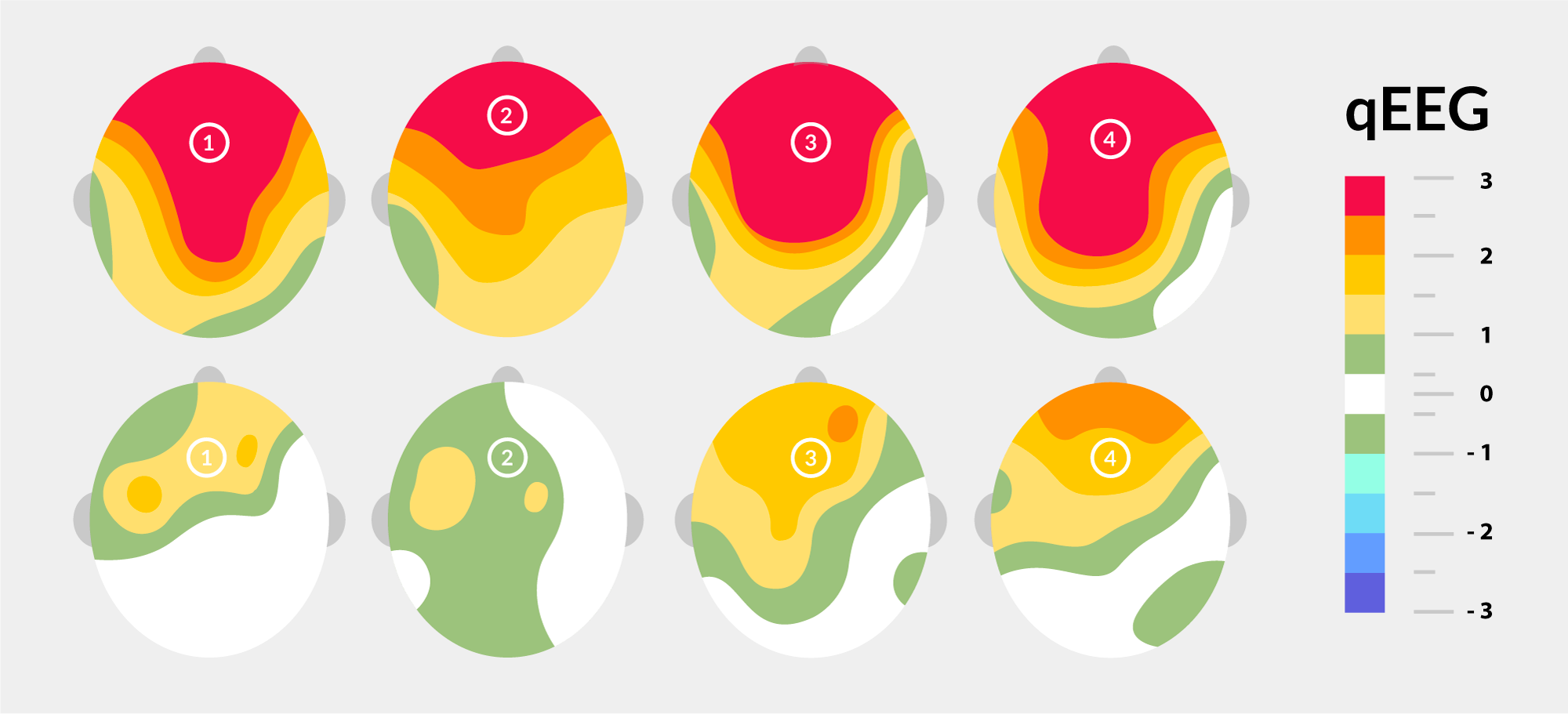
Evidence Based Practice and Research on the Efficacy of Neurostimulation and qEEG Brain Mapping
“Clinical QEEG and Neurotherapy” by Thomas F. Collura
State-of-the-art non-invasive brain–computer interface for neural rehabilitation: A review
Mental Imagery and Brain Regulation—New Links Between Psychotherapy and
The Low Energy Neurofeedback System (LENS): Theory, Background, and Introduction
EEG-Neurofeedback as a Tool to Modulate Cognition and Behavior: A Review Tutorial
PTSD Remediation with Neurofeedback
Editorial: Neuromodulation in Basic, Translational and Clinical Research in Psychiatry
Noninvasive and Invasive Neuromodulation for the Treatment of Tinnitus: An Overview
The use of EEG Biofeedback/Neurofeedback in psychiatric rehabilitation.
Revisiting the Potential of EEG Neurofeedback for Patients With Schizophrenia
Neurostimulation for Mixed Trauma Syndrome
Neuromodulatory Approaches for Depression, Adaptive Neurostimulation, and Emerging DBS Technologies
Double Trouble: Treatment Considerations for Patients with Comorbid PTSD and Depression
Neuro-stimulation Techniques for the Management of Anxiety Disorders: An Update
Peripheral nerve neurostimulation
CLINICAL TRIAL OF RESPONSIVE NEUROSTIMULATION OF THE AMYGDALA FOR TREATMENT
Defining focal brain stimulation targets for PTSD using neuroimaging
Proceedings of the 2020 ISNR Annual Conference: Keynote and Plenary Sessions
