The Subcortical Brain and the Roots of the Unconscious
Evolutionary Pathways from the Parietal Eye to Affective Consciousness
The human mind presents itself as a paradox of evolutionary engineering. Conscious awareness—that luminous theater of thought, language, and self-reflection—represents merely the visible portion of a vast cognitive architecture. Beneath the folded convolutions of the neocortex lies an ancient terrain that we share with lineages stretching back to the earliest vertebrates: the subcortical brain. This realm, comprising the brainstem, diencephalon, basal ganglia, and limbic structures, is not merely a vestigial remnant of our evolutionary past, nor a passive relay station for cortical commands. It is the metabolic and affective engine of sentience itself—the generator of the raw feeling states that depth psychology has long termed the unconscious.
This article advances a comprehensive thesis regarding the theoretical pathways of brain evolution, specifically examining the transition from external sensory orientation to internal homeostatic regulation. We investigate the hypothesis that the roots of the human unconscious are physically instantiated in the evolutionary repurposing of ancient sensory organs—most notably the parietal eye of ectothermic vertebrates—into the neuroendocrine and affective circuits of the mammalian brain. The central evolutionary mechanism proposed here is one of invagination and internalization: organs that once monitored the external environment (solar radiation, magnetic fields, physical predators) were progressively folded inward to monitor the internal milieu (circadian rhythms, somatic markers, emotional valences).
Before proceeding, we must acknowledge that this thesis operates at the intersection of established neuroscience and speculative synthesis. The evolutionary narrative presented here is neuroplausible rather than definitively proven—it represents a coherent framework for understanding clinical phenomena rather than a claim to paleontological certainty. As a clinician rather than an evolutionary biologist, I present these connections as heuristically valuable for therapeutic practice while remaining epistemically humble about their ultimate verification.
The Parietal Eye in Reptilian Ancestors
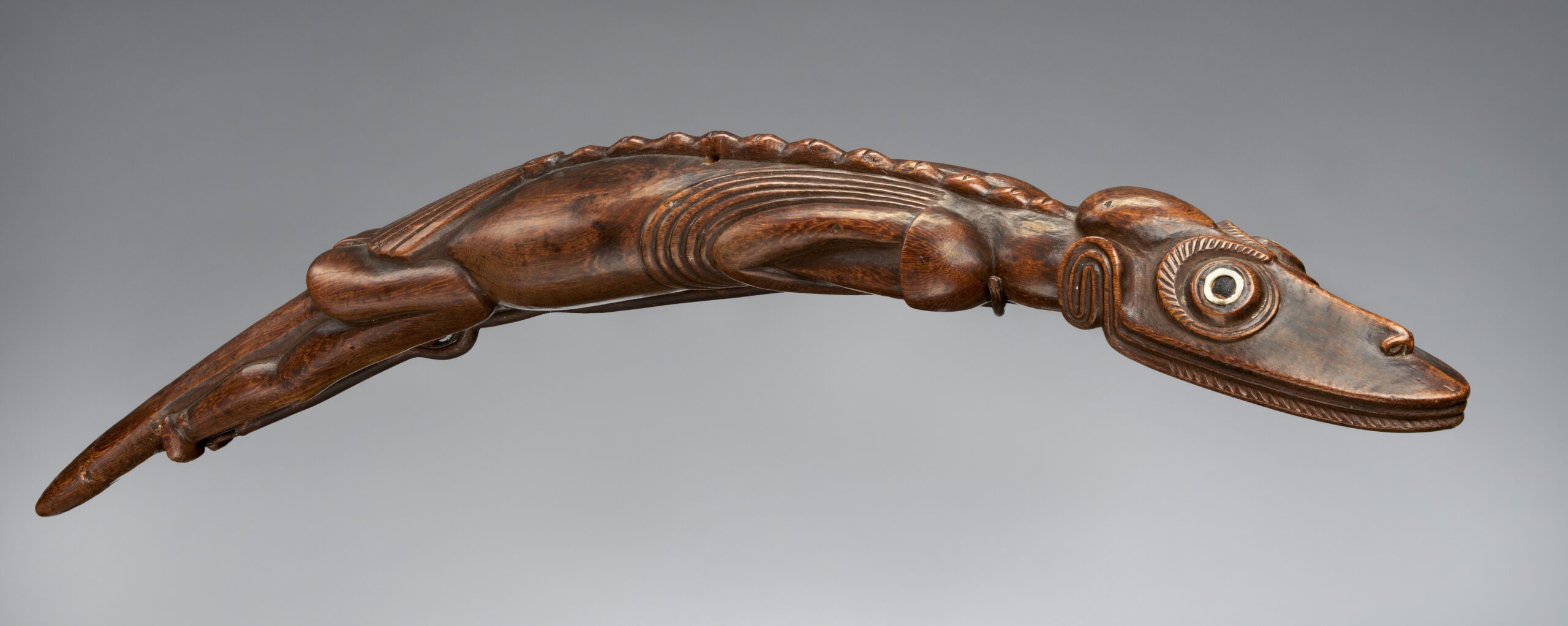
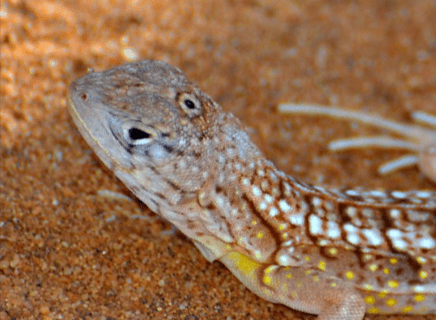 To understand the evolutionary origins of intuitive capacities and their relationship to trauma responses, we must descend into the deep time of vertebrate evolution, specifically to the sensory apparatus of the Permian and Triassic periods. Many ancient reptiles, and their modern descendants such as the tuatara (Sphenodon punctatus) and various iguanid lizards, possess a remarkable sensory organ known as the parietal eye, pineal eye, or colloquially the “third eye.”
This organ is not metaphorical but anatomically distinct—positioned on the dorsal surface of the head, typically beneath a translucent scale called the parietal scale or cornea, within an aperture in the skull roof known as the parietal foramen. Structurally, the parietal eye exhibits remarkable sophistication: it possesses a lens, a cornea, and a retina containing photoreceptor cells morphologically and biochemically similar to the cones of the lateral eyes. These photoreceptors utilize opsins—light-sensitive proteins—to transduce photon strikes into neural signals, employing the same phototransduction cascade found in the main visual system.
Functional Complexity Beyond Simple Thermometry
The primary evolutionary mandate of the parietal eye in ectotherms concerns the regulation of thermoregulation and circadian rhythms. As “cold-blooded” organisms, reptiles depend on external heat sources to facilitate metabolic reactions. The parietal eye detects the intensity, angle, and polarization of sunlight, providing the organism with critical data regarding time of day and season. This sensory input drives the pineal organ (epiphysis cerebri) and the habenula to regulate behavioral state: high solar intensity signals a metabolic window for hunting, mating, and digestion; low intensity or darkness signals the need for conservation, torpor, or shelter-seeking to prevent metabolic crash.
However, research into diverse reptilian taxa challenges the simplistic view of the parietal eye as merely a solar thermometer. Phylogenetic comparative studies of Liolaemus lizards inhabiting diverse climatic zones from sea level to high Andean elevations found that parietal eye size did not vary meaningfully with latitude, altitude, or environmental temperature, showing only weak correlation with thermal tolerance. The high intraspecific variation observed suggests that the organ’s function encompasses complex ecological adaptations related to orientation, navigation, predator detection, and precise synchronization of biological rhythms that are not yet fully understood.
A Skeptical Challenge: Is the Parietal Eye Relevant to Human Psychology?
A reductionist critique might argue: “The parietal eye is an organ found in reptiles and certain fish. Humans do not possess this structure. To draw connections between a reptilian photoreceptor and the human unconscious represents an unwarranted leap—a category error that conflates anatomical homology with functional continuity. Moreover, the ‘triune brain’ model that underlies much of this evolutionary psychology has been thoroughly criticized as oversimplified. The brain did not evolve in discrete layers like geological strata; it coevolved as an integrated system.”
This critique deserves serious engagement. The triune brain model proposed by Paul MacLean has indeed been criticized for oversimplifying a complex evolutionary process. However, the critique conflates two distinct claims: (1) that the brain evolved in discrete, encapsulated modules, and (2) that evolutionary older structures persist and influence behavior in modern organisms. The first claim is problematic; the second is well-established.
Modern evolutionary developmental biology (evo-devo) demonstrates that the basic Bauplan of the vertebrate brain—including the division into forebrain, midbrain, and hindbrain, and the differentiation of the epithalamus, thalamus, and hypothalamus—is remarkably conserved across vertebrate lineages. The pineal gland exists in all vertebrates, from lampreys to humans. The superior colliculus (optic tectum in non-mammals) retains its role in orienting responses across all vertebrate classes. The periaqueductal gray coordinates defensive behaviors in fish, amphibians, reptiles, birds, and mammals alike.
The argument is not that humans possess a literal parietal eye, but that the functional circuits served by this organ in ancestral vertebrates have been repurposed, internalized, and elaborated in the mammalian brain. This is the fundamental principle of exaptation—the co-option of existing structures for new functions—which is central to modern evolutionary theory. The cellular lineage is preserved: pinealocytes retain molecular markers of their retinal ancestry, including S-antigen (arrestin), recoverin, and the shared developmental expression of homeobox genes such as Xrx1 in both eye and pineal tissue.
The Great Internalization: From Mesothermy to Endothermy
The transition from reptilian synapsids to true mammals constitutes a metabolic revolution that fundamentally altered the architecture of the brain. The fossil record of the Therapsida—the “mammal-like reptiles” preserved in the Karoo Basin of South Africa—provides granular evidence of this transformation. Paleontological surveys of the parietal foramen across Permo-Triassic lineages reveal a gradual, convergent loss of this opening in multiple lineages leading toward mammals.
The Probainognathia, the lineage leading directly to mammaliaforms, represents the only group to achieve complete loss of the parietal foramen. This morphological closure correlates with the physiological transition from mesothermy (an intermediate metabolic state) to true endothermy (warm-bloodedness). Two factors drove this transition:
The Metabolic Driver: As early mammals evolved high basal metabolic rates, they became capable of generating their own body heat through brown adipose tissue thermogenesis and increased mitochondrial density. The reliance on the sun as an external energy source for metabolism became less critical. Consequently, the “eye” that monitored solar radiation became redundant for immediate thermal survival.
The Nocturnal Bottleneck: Many early mammals were small, nocturnal insectivores occupying ecological niches left vacant by the dominant diurnal dinosaurs. In the darkness of Mesozoic nights, a solar radiometer provided limited utility, further selecting for the closure of the parietal foramen and the reduction of direct photosensitivity.
However, evolution rarely operates through simple deletion—it is fundamentally a process of transformation and repurposing. The pineal organ did not atrophy. Instead, as the skull roof closed over it, the organ was sealed within the cranial vault. This physical internalization forced a functional inversion: the organ that once looked outward to synchronize the organism with the world began to look inward to synchronize the organism’s internal systems.
Cellular Metamorphosis: The Photoneuroendocrine Lineage
The “switch” from sensory organ to secretory gland is preserved in the cellular biology of the pineal. In non-mammalian vertebrates, pineal cells are direct photoreceptors. In birds and some reptiles, they are “modified photoreceptors” that retain light sensitivity while also engaging in secretory activity. In mammals, these cells have transformed into pinealocytes—specialized secretory cells that produce melatonin in response to neural rather than photic input.
Despite this transformation, pinealocytes retain the molecular “memory” of their retinal ancestors. They share a conserved signaling pathway known as the tryptophan triad and express specific proteins critical for phototransduction in the eyes. Genetic studies in Xenopus reveal that the homeobox gene Xrx1 is expressed during development of both eye and pineal gland, underscoring their shared origin from the neural ectoderm—the same embryonic tissue that gives rise to the entire nervous system.
The mammalian pineal gland thus functions as a “folded-in” retina. It receives information about the light-dark cycle not through a hole in the skull, but via a circuitous neural pathway: Light → Retinal Ganglion Cells → Retinohypothalamic Tract → Suprachiasmatic Nucleus (SCN) → Paraventricular Nucleus (PVN) → Superior Cervical Ganglion → Pineal Gland. The external “god” (the sun) has been replaced by a neural signal (norepinephrine), which triggers pinealocytes to synthesize melatonin in the absence of light.
This represents the first theoretical pathway of the “unconscious”: Exteroception (Sensory) → Interoception (Regulatory). The brain ceased looking solely outward for guidance and began to synthesize an internal model of time—a stable temporal “self” that persists regardless of external lighting conditions.
Recapitulation: The Ghost of the Third Eye in the Neonate
A striking piece of evidence supporting this evolutionary narrative is found in the ontogeny of the mammalian pineal gland. In developmental biology, the principle that “ontogeny recapitulates phylogeny” often holds true for conserved structures. Research into neonatal rats has revealed that for a transient period after birth (up to 17 days), pinealocytes exhibit morphological differentiation remarkably similar to retinal photoreceptors. Electron microscopy reveals the formation of ciliary structures with lamellated and vesicular membranes, mimicking the outer segments of rods and cones. Furthermore, these developing pineal cells express a suite of genes involved in the phototransduction cascade, including rhodopsin kinase, arrestin, and cyclic GMP-gated channels.
This “transient photoreceptor differentiation” suggests that the genetic program for the “third eye” is still encoded in the mammalian genome. It is expressed briefly during early development before being suppressed by the maturation programs that enforce a secretory fate. This phenomenon indicates that the pineal gland is not merely a gland but a latent eye—an organ that has been folded inward and blinded to the outside world, yet remains chemically responsive to the memory of light.
This biological fact serves as a potent theoretical bridge. If the pineal gland retains the “memory” of being an eye, does the brain retain the “memory” of the orientation it once provided? The hypothesis advanced here is that the functions of the parietal eye—orientation, threat detection, and circadian alignment—were not lost but were distributed to other subcortical structures, forming the physiological basis of the unconscious mind.
The Retinal Shift: ipRGCs as the Internalized Sun
The “Switch”: From Parietal to Retinal Dominance
With the involution of the parietal eye, the mammalian brain faced a critical engineering problem: How to maintain synchronization with the solar day without a dedicated solar sensor? The lateral eyes, evolved for high-acuity image formation, were ill-suited for this task. Rods and cones adapt rapidly to changing light levels, optimizing for contrast rather than absolute irradiance. A cloud passing over the sun should not reset the biological clock.
The evolutionary solution was the emergence of a third class of photoreceptor within the retina itself: the Intrinsically Photosensitive Retinal Ganglion Cells (ipRGCs). Unlike rods and cones, which are ciliary photoreceptors, ipRGCs utilize melanopsin (Opn4), a photopigment evolutionarily related to the rhabdomeric opsins found in the eyes of invertebrates (like flies and octopuses) and, crucially, in the parietal eyes of lower vertebrates.
This suggests a profound evolutionary conservation. The chemical system used by the “third eye” to detect irradiance was not discarded; it was migrated into the ganglion cell layer of the lateral eyes. This “switch” effectively embedded the function of the parietal eye within the visual eye, creating a duplex retina where “conscious” vision (images) and “unconscious” vision (irradiance/time) operate in parallel but distinct pathways.
ipRGCs: The Sluggish Counters of Photons
The physiology of ipRGCs reflects their role as the successor to the parietal eye. They are relatively insensitive to light compared to rods and cones, requiring high-intensity illumination to activate. Their response is “sluggish”: they depolarize slowly upon light onset and continue to fire for a long duration after the light is extinguished. They integrate photon flux over time and space, blurring temporal and spatial details to provide a stable measurement of ambient light intensity.
This response profile is practically useless for seeing a predator or finding food (image forming). However, it is perfect for measuring the length of the day (circadian entrainment) and the brightness of the environment (pupillary reflex). This functional dichotomy—fast/detailed versus slow/integrating—mirrors the distinction between the “Ego” (conscious, detail-oriented processing) and the “Id” or “Unconscious” (slow, state-oriented regulation).
The Projections of the “Shadow Eye”
The axons of ipRGCs form the retinohypothalamic tract, but their targets extend far beyond the circadian clock. While the Suprachiasmatic Nucleus (SCN) is the primary target for circadian synchronization, ipRGCs innervate dozens of subcortical targets that have little to do with image formation and everything to do with “state” and “affect”:
The Suprachiasmatic Nucleus (SCN): The master clock.
The Olivary Pretectal Nucleus (OPN): Controlling the pupillary light reflex.
The Ventrolateral Preoptic Area (VLPO): A sleep-promoting center.
The Lateral Habenula (LHb) and Perihabenular Nucleus (pHb): Regulating mood, reward, and the “anti-reward” system.
The Medial Amygdala: Involved in innate fear and social behavior.
The Superior Colliculus: Involved in orienting and threat detection.
The M4 Subtype and the Mood Pathway
Recent research has identified distinct subtypes of ipRGCs (M1–M6) with specific projection patterns. Of particular interest to the thesis of the “unconscious” are the M4 ipRGCs. These cells have been shown to project directly to the Lateral Habenula (LHb) via the ventral Lateral Geniculate Nucleus (vLGN) and Intergeniculate Leaflet (IGL).
The LHb is a critical node in the brain’s emotional circuitry. The direct projection from the retina to the habenula means that environmental light can influence mood and motivation independently of the image-forming visual cortex and even independently of the circadian clock. This pathway allows light to act as a direct modulator of the “anti-reward” system.
When we speak of “seasonal depression” or the “mood-lifting” effect of sunlight, we are describing the activity of this pathway. It is a non-visual, non-conscious channel through which the environment regulates the “inner weather” of the psyche. This is the functional internalization of the parietal eye: the sun is no longer just a compass for navigation; it is a lever for emotional regulation.
The Lateral Habenula: Gatekeeper of Darkness and the “Shadow”
Anatomy of an Ancient Monitor
The habenula is a phylogenetically ancient structure located in the epithalamus, physically adjacent to the pineal gland. Conserved across all vertebrates from lampreys to humans, it serves as a critical link between the forebrain (limbic system, basal ganglia) and the midbrain (dopaminergic and serotonergic nuclei).
In many lower vertebrates, the habenula shows marked asymmetry, often correlated with the asymmetry of the parietal eye/pineal complex. In mammals, this asymmetry is reduced, but the functional division between the Medial Habenula (MHb) and Lateral Habenula (LHb) remains distinct. The LHb, in particular, has emerged as a central player in the understanding of depression, aversion, and negative reinforcement.
The “Anti-Reward” Center
While the dopamine system of the Ventral Tegmental Area (VTA) and Nucleus Accumbens mediates reward and desire (Panksepp’s “SEEKING” system), the Lateral Habenula functions as the “Anti-Reward” center. The LHb is activated by aversive stimuli (pain, stress, bitter taste), the omission of an expected reward (disappointment), and cues predicting punishment.
When the LHb fires, it sends powerful excitatory (glutamatergic) signals to the Rostromedial Tegmental Nucleus (RMTg), also known as the “tail of the VTA.” The RMTg is a GABAergic brake; when activated by the habenula, it inhibits the dopamine neurons of the VTA and the serotonin neurons of the Dorsal Raphe Nucleus (DRN).
The Equation of Misery: LHb Activation → RMTg Activation → VTA/Raphe Inhibition → Drop in Dopamine/Serotonin → Anhedonia, Passive Coping, “Giving Up.”
This mechanism is evolutionarily adaptive. It teaches the organism to stop pursuing fruitless goals, to withdraw from dangerous environments, and to conserve energy when resources are scarce. However, when this system becomes hyperactive or tonically active, it results in the phenomenology of Major Depressive Disorder: a persistent state of “anti-reward,” where nothing feels good and the future looks bleak.
The “Darkness” Mechanism: A Photo-Inhibitory Circuit
The connection between the ipRGCs and the LHb provides a stunning insight into the biological roots of “darkness” as a psychological state. Research indicates that light exposure—specifically the blue light detected by melanopsin—inhibits the firing of Lateral Habenula neurons.
Light Presence: ipRGCs activate inhibitory interneurons (likely in the vLGN/IGL or pHb) that suppress the LHb. Result: The “brake” on dopamine is lifted. Motivation and mood are sustained.
Light Absence (Darkness): The inhibitory drive on the LHb is removed. The LHb becomes active (or disinhibited). Result: The “brake” is applied to dopamine/serotonin. The organism shifts into a state of withdrawal, energy conservation, and lowered mood.
This is the “Hibernation” hypothesis of depression. In our evolutionary past, darkness (winter) signaled a lack of resources and a need for dormancy. The “Darkness” signal, mediated by the activation of the habenula, triggered a metabolic and behavioral shutdown. In modern humans, this ancient circuit manifests as Seasonal Affective Disorder (SAD) or non-seasonal depression when we are deprived of high-intensity light signals.
This mechanism explains why “Darkness” is the universal archetype of the Shadow, fear, and depression. It is not a metaphor. It is a direct neurophysiological state. Darkness literally activates the brain’s punishment center. The “Shadow” of Jungian psychology has a physical address in the epithalamus.
The Perihabenular Nucleus (pHb): The Mood-Light Transducer
Recent studies identify the Perihabenular Nucleus (pHb), a distinct region of the dorsal thalamus, as a key mediator of light’s effect on mood. The pHb receives direct innervation from M1 and M4 ipRGCs and projects to the Ventromedial Prefrontal Cortex (vmPFC) and the Nucleus Accumbens.
The pHb appears to function as a dedicated channel for “mood-light” integration, separate from the “clock-light” integration of the SCN. This segregation ensures that light can influence affect independently of circadian phase. Chronic activation of the pHb by aberrant light cycles (e.g., light at night) has been shown to induce depressive-like behaviors, suggesting that “light pollution” acts as a stressor that dysregulates this ancient orientation system.
The Subplate Zone: The Transient Scaffold of the Mind
The Expansion of the Subplate in Human Evolution
While the habenula and pineal represent the conservation of the ancient, the mammalian neocortex represents the explosion of the new. However, the construction of this massive neocortex relies on a “transient” structure that mirrors the transient photoreceptors of the pineal: the Subplate Zone.
The subplate is a layer of neurons that appears early in fetal development, situated beneath the cortical plate. In rodents, it is a thin band. In primates and humans, it is massively expanded, becoming the thickest zone of the fetal telencephalon. It serves as a “waiting compartment” for thalamocortical afferents—axons from the thalamus carrying sensory information from the eyes, ears, and skin arrive at the cortex weeks before the cortical neurons (layer IV) are ready to receive them. They “wait” in the subplate, forming functional synapses with subplate neurons.
The “Internalization” of Developmental Plasticity
Kostovic and Rakic have highlighted that the human subplate is not merely a waiting room but a site of sophisticated synaptic activity. Subplate neurons are the first generated neurons of the cerebral cortex and are responsible for the initial processing of sensory input in the fetus and preterm infant. They drive the spontaneous activity (e.g., retinal waves) that sets up the columnar architecture of the cortex before the eyes even open.
Crucially, the majority of subplate neurons are programmed to die (apoptosis) or dissolve into the interstitial neurons of the white matter once the permanent cortical circuits are established. This transient existence parallels the involution of the parietal eye. Just as the third eye provided a scaffold for physical orientation that was later internalized, the subplate provides a scaffold for cognitive orientation that is “dissolved” into the unconscious infrastructure of the white matter.
The Subplate as the “Ghost” in the Machine
The survival of a subset of subplate neurons as Interstitial Neurons of the White Matter suggests that they may retain a role in modulating cortical activity from the “shadows.” These neurons are chemically diverse (GABAergic, glutamatergic, peptidergic) and are strategically positioned to monitor the traffic between the thalamus and the cortex.
If the subplate “trains” the cortex using ancient thalamic inputs, it may be the conduit through which subcortical “archetypal” structures imprint themselves onto the developing neocortex. The “tabula rasa” of the cortex is not blank; it is pre-patterned by the subplate. The “transient” neurons that die off leave behind a “ghost” architecture—the functional connectivity maps that define how we perceive the world. This aligns with the Jungian notion that the collective unconscious is “inherited” structure—the subplate may be the mechanism of that inheritance, translating the genetic code of the “Old Brain” into the wiring of the “New Brain.”
The Pineal Gland: Valve of the Chemical Mind
If the parietal eye was the sensor of the external world, the mammalian pineal gland is the regulator of the internal world. Its role extends beyond simple hormone secretion; it functions as a transducer that converts neural signals from the sympathetic nervous system into chemical outputs that modulate the entire brain-body axis, effectively establishing the “state” of the unconscious.
Melatonin and the Biochemistry of Darkness
The primary output of the pineal gland is N-acetyl-5-methoxytryptamine, commonly known as melatonin. This hormone is synthesized from serotonin—a neurotransmitter critical for mood and waking consciousness—in a rhythm strictly antiphasic to daylight. The synthesis pathway is controlled by the rate-limiting enzyme arylalkylamine N-acetyltransferase (AANAT), which is suppressed by light exposure via the retinohypothalamic pathway.
In the context of depth psychology, melatonin acts as the chemical messenger of the Shadow. When the conscious mind (associated with the solar/cortical waking state and serotonin dominance) recedes, melatonin levels rise, ushering in the domain of the unconscious—sleep, dreams, and restoration. This is not merely metaphor: melatonin modulates the activity of the reticular activating system and the thalamus, effectively “closing the gates” to sensory input and allowing endogenously generated content—dreams and unconscious material—to dominate neural activity.
The pineal gland’s production of melatonin is a vestige of its ancient role. In early life forms, melatonin likely evolved as an antioxidant to protect against oxidative stress caused by high ultraviolet exposure (detected by the parietal eye). In mammals, this protective function was co-opted into a signaling function. The “chemical darkness” produced by the pineal signals the entire organism to shift into a restorative, anabolic state, mimicking the torpor that reptiles entered when the sun descended.
Sensory Gating: The Biological Filter Between Worlds
A critical theoretical link between the pineal gland and the structure of the human psyche is its influence on sensory gating. Sensory gating is the brain’s neurological capacity to filter out redundant or irrelevant stimuli from the environment, preventing information overload and maintaining coherent perception. This function is largely orchestrated by the thalamus, often described as the “gateway to the cortex.”
Research indicates that the pineal gland, via melatonin and its precursors, modulates the excitability of thalamic relay nuclei. By influencing the firing patterns of thalamic neurons—shifting them from “tonic” mode (faithful transmission of sensory data) to “burst” mode (disconnection from sensory input)—melatonin helps maintain the boundary between internal and external worlds. The thalamic reticular nucleus, in particular, serves as a kind of attentional “spotlight” that determines which sensory streams reach cortical awareness.
The P50 auditory evoked potential is a standard electrophysiological measure of this sensory gating. In healthy individuals, if two identical clicks are presented in rapid succession (typically 500ms apart), the brain suppresses its response to the second click—it “gates it out” as redundant. In conditions like schizophrenia, this P50 suppression is often defective. The patient’s brain fails to filter the redundant stimulus, leading to a flooding of conscious awareness with sensory debris and internal associations—a state frequently described as an invasion of the conscious mind by unconscious material.
Skeptical Challenge: Is Sensory Gating Really Related to the Unconscious?
A cognitive neuroscientist might object: “Sensory gating is an attentional mechanism, not a gateway to the ‘unconscious.’ The concept of the unconscious is a psychoanalytic construct that has no clear neurobiological referent. What you’re describing is simply information filtering—a basic computational function that has nothing to do with repressed memories, archetypes, or Jungian complexes. To conflate thalamic gating with depth psychology represents a categorical confusion between mechanism and meaning.”
This objection reflects a legitimate concern about reification. However, it rests on an impoverished understanding of what “unconscious” means in contemporary neuroscience and depth psychology. The unconscious is not merely a repository of repressed memories—it encompasses the vast majority of neural processing that occurs below the threshold of awareness, including:
Implicit memory systems (procedural learning, emotional conditioning), autonomic regulation (heart rate variability, respiratory patterns, visceral signaling), affective states generated by subcortical circuits (Panksepp’s SEEKING, RAGE, FEAR, PANIC, CARE, PLAY, and LUST systems), and predictive models maintained by the brain that never reach awareness but shape perception and action.
When sensory gating fails, these normally subthreshold processes—affect, association, implicit memory—intrude into awareness. The patient with schizophrenia does not simply experience “too much sensory input”; they experience a breakdown in the boundary between self-generated and world-generated content. Internal speech becomes external hallucination. Implicit associations become paranoid convictions. The subjective experience is precisely one of the unconscious erupting into consciousness.
Mark Solms’ neuropsychoanalytic research provides crucial support here. Solms argues, contra Freud, that the Id (subcortical affect systems) is inherently conscious—it feels rage, fear, desire. What is “unconscious” is often the cortex’s defensive management of these affects. The thalamic gating system is thus not merely filtering “information” but regulating the boundary between primary affective consciousness (subcortical) and secondary representational consciousness (cortical). When gating fails, the raw affect of the Id floods the representational systems of the Ego.
The Dual Nature of Intuition and Trauma
As evolution progressed and the parietal eye regressed in early mammals, the pineal gland and its deep connections to the limbic system and subcortical brain took on new functions and significance. While the pineal gland lost its direct photosensitivity, it retained a key role in regulating circadian rhythms, sleep-wake cycles, and states of consciousness through melatonin secretion.
However, the pineal gland’s influence extends beyond mere physiological regulation. Situated as a nexus between ancient reptilian brain structures and more recently evolved limbic and neocortical regions, the pineal gland and its associated networks serve as a “primal antenna” for subtle environmental and internal cues. This deep, embodied wisdom of the pineal-limbic system often manifests as intuitive “gut feelings,” “hunches,” or instinctive responses that arise from beyond conscious thought.
Interestingly, this intuitive mode of knowing shares qualities with the spatial awareness functions of the parietal eye in lower vertebrates. Just as the parietal eye provided a direct, non-visual pathway for detecting changes in light, movement, and orientation, the pineal-limbic system offers a “felt sense” of the world—an immediate, pre-verbal attunement to the energetic and emotional landscape within and around us. The situational awareness capacities mediated by the parietal eye have been internalized and transformed into a more abstract, intuitive form of perception.
This transition reflects the larger shift from the concrete, sensorimotor cognition of early vertebrate ancestors to the more symbolic, conceptual cognition of the human mind. As the parietal eye atrophied and its functions were subsumed by deeper brain structures like the superior colliculus and posterior parietal cortex, the raw data of sensory perception was increasingly filtered through layers of associative memory, emotional valence, and narrative meaning.
The result is a kind of “mapping” of the external world onto the internal landscape of the psyche—a projection of our unconscious contents and complexes onto the screen of reality. In this way, the intuitive wisdom of the pineal-limbic system can be both a source of profound insight and a potential trap, leading us to mistake our own unresolved fears, desires, and traumas for objective truth.
The Shadow Side: When Intuition Becomes Pathology
On one hand, the pineal-limbic system and its associated networks are the wellspring of our deepest creativity, empathy, and spiritual connection. When this system is functioning optimally, we have a strong sense of attunement to ourselves, others, and the world. We can access a kind of “direct knowing” that bypasses the discursive intellect and speaks in the language of symbol, metaphor, and felt meaning.
On the other hand, this same system is the seat of our most primal wounds and reactive patterns. When the limbic system and brainstem are overwhelmed by traumatic stress, they can become chronically hyperaroused or dissociated, leading to dysregulation and disconnection from body and environment. In this state, the individual may feel trapped in “survival mode,” constantly scanning for threats and unable to access higher-order capacities for reasoning, perspective-taking, and self-reflection.
This is where Carl Jung’s concept of the Shadow becomes particularly relevant. For Jung, the Shadow represents the repressed, rejected, or unconscious aspects of personality that are split off from the conscious ego and projected onto the external world. These shadow contents are often rooted in early experiences of trauma, neglect, or overwhelming emotion—experiences too painful or threatening to integrate into our conscious self-image.
When possessed by a complex or traumatic shadow, we may find ourselves repeatedly drawn into destructive patterns of thought and behavior, as if caught in the gravitational pull of a black hole. We may feel deep shame, worthlessness, or fear that colors all experiences and relationships. Critically, we may mistake the voice of the wounded shadow for the voice of intuitive wisdom, making choices that perpetuate suffering.
The task of healing and integration is to bring these shadow contents into the light of conscious awareness, so they can be met with compassion, understanding, and choice. This is the essence of Jung’s individuation process—the lifelong journey of becoming more fully ourselves by embracing and integrating all of our disparate parts and potentials.
Magnetoreception: The Sixth Sense and the Physics of Intuition
A key component of the subcortical thesis is the existence of sensory modalities that operate below the threshold of conscious awareness, feeding into what is colloquially termed “intuition.” Magnetoreception—the ability to detect Earth’s geomagnetic field—is well-established in birds, turtles, insects, and fish, where it is utilized for navigation. Recent evidence suggests this sense is also present, if latent, in humans, mediated by the very same cryptochromes found in the pineal-retinal complex.
Cryptochromes and the Radical Pair Mechanism
The leading theoretical model for magnetoreception is the Radical Pair Mechanism within Cryptochromes. Cryptochromes are flavoproteins found in the retina and other tissues that are sensitive to blue light and serve as evolutionarily conserved regulators of the circadian clock. In migratory birds, CRY1a in the retina is believed to function as the primary magnetosensor.
The mechanism is genuinely quantum biological: Blue light strikes the flavin cofactor in the cryptochrome, triggering electron transfer that creates a “radical pair”—two molecules with unpaired electrons that are spatially separated but quantum entangled. The spin state of these electrons (singlet versus triplet) acts as a delicate chemical balance. Earth’s magnetic field, though weak (approximately 25-65 microtesla), is sufficient to influence the oscillation between these spin states. The ratio of singlet to triplet states determines the yield of the signaling state of the protein, which in birds theoretically produces a visual modulation—the magnetic field is “seen” as a pattern of brightness or color superimposed on the visual field.
Human Magnetoreception: Alpha-ERD and Subconscious Processing
For decades, human magnetoreception was dismissed as pseudoscience. However, groundbreaking research by the Caltech Human Magnetic Reception Laboratory (Kirschvink et al., 2019) demonstrated that humans respond to magnetic fields neurophysiologically, even without conscious awareness.
Using high-density EEG in a Faraday cage, researchers exposed subjects to rotating magnetic fields matching Earth’s geomagnetic field strength. They observed a distinct, repeatable drop in alpha-wave amplitude (Alpha-Event Related Desynchronization, or α-ERD) in parietal and occipital regions. Alpha desynchronization is a standard marker of sensory processing and attention—it indicates that the brain is “waking up” to a stimulus.
Crucially, subjects reported feeling nothing. The brain was “noticing” the magnetic shift, processing it in parietal and occipital cortex, and reacting to it, yet this data was not promoted to the “Global Workspace” of consciousness. This provides hard biological evidence for unconscious perception: the brain possesses a data stream regarding environmental orientation that it processes subcortically or in early cortical areas but which does not enter conscious awareness.
Skeptical Challenge: Is Human Magnetoreception Functionally Meaningful?
A skeptic might argue: “Even if humans show neural responses to magnetic fields, this doesn’t mean the sense is functionally meaningful. Migratory birds use magnetoreception for navigation—they fly thousands of miles guided by magnetic maps. Humans don’t migrate; we use GPS. An atavistic sensitivity to magnetic fields, even if present, would be like the appendix—a vestigial structure with no modern function. To claim this as a basis for ‘intuition’ is romantic speculation without empirical support.”
This critique requires engagement with recent behavioral data. Research on the game of Go demonstrated that players’ decisions were influenced by artificial magnetic fields. Specifically, the geomagnetic field affected subconscious binary decision-making (the initial intuitive choice) but not conscious, intentional verification. This suggests that magnetic fields influence the “gut” or “initial impulse” layer of cognition—precisely the level associated with intuition.
Furthermore, the argument need not rest on magnetoreception for navigation to be meaningful. Evolution “switched” explicit navigation (knowing which way is North for migration) for implicit orientation (feeling “right” versus “wrong” in an environment). The signal from retinal cryptochromes—human hCRY2 has been shown to be magnetosensitive when expressed in transgenic Drosophila—likely feeds into the hypothalamus and limbic system rather than visual cortex. This produces a “somatic marker” or “affective tone” associated with a place or direction, rather than a visual compass.
This aligns with the Jungian concept of the unconscious as a source of “knowledge” that the ego does not possess. The parietal eye function of orientation has not disappeared—it has been distributed into the retina and the subconscious processing loops of the brain. When people report “bad feelings” about places or situations, they may be processing genuine environmental data that never reaches explicit awareness.
The Sentinel of the Unconscious: The Superior Colliculus
While the pineal gland regulates the timing of the unconscious, the Superior Colliculus serves as its sentinel. Located in the midbrain tectum (the “roof”), the SC is the mammalian homolog of the optic tectum, which is the primary visual processor in non-mammalian vertebrates. It represents the persistence of the “reptilian visual brain” beneath the mammalian neocortex.
The Two Visual Systems: Ancient and Modern
The human visual system is dualistic, reflecting the evolutionary transition from reptile to mammal:
The Geniculostriate Pathway (“New” Way): Retina → Lateral Geniculate Nucleus (LGN) → Primary Visual Cortex (V1). This pathway provides conscious, high-resolution, color vision and object recognition. It is the vision of the ego—the representational, symbolic processing that identifies “what” is being seen.
The Retinotectal Pathway (“Old” Way): Retina → Superior Colliculus → Pulvinar → Amygdala/Parietal Cortex. This pathway operates extremely fast (latency < 20-30ms) and completely unconsciously. It is the vision of the Id—the affective, survival-oriented processing that determines “where” and “how threatening.”
The SC does not “see” faces or colors in high fidelity; it detects salience: looming shadows, rapid movement, the coarse configuration of eyes and faces. Its primary function is the Orienting Reflex—the rapid, involuntary alignment of eyes, head, and body toward a novel or potentially threatening stimulus.
Blindsight: Proof of the Unconscious Visual Brain
The existence of the unconscious visual brain is most dramatically demonstrated by the phenomenon of blindsight. Patients with destruction of the primary visual cortex (V1) are clinically blind—they report seeing nothing in the affected visual field. However, if a ball is thrown at them, they can catch it. If shown a fearful face, their amygdala activates and they may report feeling “uneasy” without seeing the face. They report “sensing” something without “seeing” it.
This ability is mediated by the subcortical pathway: Retina → Superior Colliculus → Pulvinar → Amygdala. This “Low Road” bypasses the cortex entirely. It allows for the rapid detection of threat (e.g., a snake, a fearful face) and constitutes the biological basis for “gut feelings” or intuition—perception that occurs without conscious visual representation. The SC is essentially an unconscious scanner of the environment, constantly monitoring for danger and orienting the organism’s attention.
This phenomenon confirms that visual processing and emotional evaluation occur in the subcortical brain independent of conscious awareness. The SC acts as a “threat detection interface” that scans the environment for danger and primes the amygdala before the conscious mind has even registered the image. The patient feels threat without seeing it.
Neuroception: The Subcortical Assessment of Safety
Stephen Porges coined the term “Neuroception” to describe the neural process of distinguishing safe from dangerous contexts without conscious awareness. Neuroception relies on these ancient subcortical structures (SC, periaqueductal gray, temporal cortex) to evaluate biological movements and environmental cues.
When neuroception detects Safety (Ventral Vagal), the social engagement system is active. The vagus nerve brakes the heart, promoting calm and connection. When Danger is detected (Sympathetic), the SC and amygdala trigger the sympathetic nervous system (fight/flight). When there is Life Threat (Dorsal Vagal), if the threat is overwhelming or inescapable, the ancient “reptilian” vagal circuit initiates immobilization or “shutdown” (feigning death).
The Superior Colliculus is integral to this system, as it mediates the initial orienting to the threat. Porges argues that neuroception is a “subconscious system for detecting threats and safety” that precedes and dictates our conscious emotional state.
Trauma and the Frozen Sentinel
In the context of trauma, the orienting function of the SC becomes dysregulated. The “innate alarm system,” centered on the SC and its connections to the periaqueductal gray and amygdala, becomes hypersensitive or locked. The evolutionary implication is profound: Trauma is often a failure of the Orienting Response. The reptilian brain initiates a reflex to look at the threat, but if the threat is overwhelming, the sequence is arrested. The “switch” to cortical control fails, and the organism is left in a state of subcortical looping.
This understanding has profound implications for trauma therapy. Approaches like Brainspotting and EMDR appear to work by re-engaging this arrested orienting response. By holding a fixed eye position (Brainspotting) or using lateral eye movements (EMDR), the therapist engages the retinotectal pathway, unlocking the SC from its frozen state and allowing the completion of the defensive response that was interrupted during trauma.
The Polyvagal Hierarchy: An Architecture of Defense
The architecture of the subcortical brain is further elucidated by Polyvagal Theory, developed by Stephen Porges. This theory maps the autonomic nervous system onto evolutionary time, creating a hierarchy of defense that mirrors the layers of the unconscious.
Three Phylogenetic Circuits
Porges identifies three evolutionary stages of the vagus nerve and autonomic nervous system, which function as distinct “platforms” for behavior:
Dorsal Vagal Complex (DVC): The oldest system, utilizing unmyelinated fibers originating in the Dorsal Motor Nucleus of the Vagus. Its primary function is regulation of subdiaphragmatic organs (digestion). Under life threat, it triggers immobilization (death feigning, syncope, bradycardia). This is the “Freeze” response—a metabolic shutdown. It corresponds evolutionarily to the reptilian state of torpor.
Sympathetic Nervous System (SNS): The system of mobilization. It prepares the organism for “Fight or Flight” by increasing heart rate and shunting blood to skeletal muscles. It represents an active defense strategy and corresponds to the aroused, vigilant state of early mammals.
Ventral Vagal Complex (VVC): The newest system, unique to mammals, utilizing myelinated fibers originating in the Nucleus Ambiguus. It controls the striated muscles of face, larynx, and pharynx, and acts as a “brake” on the heart. This is the Social Engagement System—it promotes safety, connection, and calm through facial expression, vocalization, and listening.
Jacksonian Dissolution: The Regression of the Self
Porges argues that under stress, the brain undergoes Jacksonian Dissolution—it dissolves from the newest, most complex systems back to the oldest, in reverse evolutionary order:
Stage 1 (Safety): The VVC is active. We use facial expressions, prosody, and eye contact to negotiate safety. The cortex is online and integrated.
Stage 2 (Danger): The Vagal Brake releases. The SNS takes over. We mobilize to fight or flee. The limbic system dominates. Cortical integration begins to fragment.
Stage 3 (Life Threat): The SNS is overwhelmed. The DVC takes over. We collapse, dissociate, or freeze. The reptilian brainstem dominates. This is the state of trauma—a regression to the most primitive survival strategy.
Skeptical Challenge: Is Polyvagal Theory Scientifically Sound?
Polyvagal Theory has faced significant academic criticism. Critics argue that the phylogenetic claims are oversimplified—the vagus nerve in fish is already complex, and the “dorsal” versus “ventral” distinction does not map neatly onto evolutionary stages. Furthermore, the claim that the dorsal vagal complex mediates “freeze” has been challenged; tonic immobility appears to involve multiple systems including the periaqueductal gray.
These critiques are valid at the level of detail. However, they do not undermine the core clinical utility of the model. What Polyvagal Theory provides is a functional taxonomy of autonomic states that maps onto subjective experience and therapeutic intervention. Whether the precise neuroanatomy is exactly as Porges describes is less important than the clinical observation that:
(1) States of social engagement, mobilization, and immobilization are phenomenologically distinct; (2) These states can become “stuck” in trauma survivors; (3) Therapeutic interventions that work with autonomic state (breath, movement, co-regulation) are clinically effective.
The model’s value is heuristic rather than definitive. Like the triune brain model, it provides a useful scaffold for understanding the hierarchical organization of defense—even if the underlying neurobiology is more complex than the model suggests.
Therapeutic Implications: Engaging the Primal Brain
The understanding that trauma is encoded in subcortical structures—and that intuition and traumatic reactivity share neural substrates—has profound implications for psychotherapy. Approaches that work directly with the body and the non-verbal mind may access and transform patterns inaccessible to purely cognitive interventions.
Somatic Experiencing
Developed by Peter Levine, Somatic Experiencing is a body-oriented approach to healing trauma that draws on insights from neuroscience, ethology, and indigenous healing practices. The core idea is that trauma is primarily a physiological phenomenon—a disruption of the natural rhythms and responses of the autonomic nervous system.
When we experience a traumatic event, our bodies mobilize massive energy to defend against threat. However, if this energy is not fully discharged, it can become “stuck” in the nervous system, leading to hypervigilance, dissociation, and emotional reactivity. SE works by gently guiding the client’s attention to the felt sense of the body, particularly to subtle sensations and impulses arising in response to trauma-related cues. By staying with these sensations and allowing them to unfold, the client can gradually release trapped energy and restore equilibrium.
The principles of SE reflect an intuitive understanding of the evolutionary role of the subcortical brain: the emphasis on instinctive, sensorimotor responses; attention to subtle shifts in arousal; trust in the body’s innate wisdom. SE can be understood as “reverse engineering” ancient survival responses, using the language of sensation and movement to communicate with primal brain patterns.
EMDR (Eye Movement Desensitization and Reprocessing)
Developed by Francine Shapiro, EMDR involves guiding the client through lateral eye movements or other bilateral stimulation while simultaneously holding the memory or image of a traumatic event. The exact mechanisms remain debated, but one theory is that bilateral stimulation helps “unfreeze” traumatic memory from its isolated, fragmented state in the subcortical brain, allowing reprocessing and integration with broader life context.
Research suggests that eye movements may target the superior colliculus and its connections to the mediodorsal thalamus. This pathway has been shown in murine models to suppress activity of the basolateral amygdala, effectively downregulating fear response and allowing cortical reintegration of traumatic memory.
Notably, the use of rhythmic, alternating stimulation echoes primal, cyclical rhythms of the natural world—the oscillation of day and night, ebb and flow of tides, contraction and relaxation of breath. By entraining the brain to these ancient evolutionary rhythms, EMDR may tap into deep regenerative capacities of the nervous system that evolved before language or rational cognition.
Brainspotting
Developed by David Grand, Brainspotting combines elements of EMDR, somatic experiencing, and mindfulness. The approach involves identifying specific eye positions or “brainspots” that activate or resonate with traumatic memories or emotional states. By holding these brainspots with focused attention and mindful awareness, the client can gently process and release underlying neurophysiological patterns.
Grand theorizes that trauma is stored in “capsules” within the subcortical brain. By holding a fixed eye position, the therapist engages the retinotectal pathway, locking the SC onto the neural coordinates of the trauma. This bypasses cortical “narrative” and accesses implicit somatic memory directly. The SC’s connectivity to the periaqueductal gray and amygdala allows processing of the “frozen” orienting response.
One possible explanation for Brainspotting’s effectiveness is that specific eye positions correspond to different “fields” of subcortical activation, reflecting the evolutionary layering of the brain. A brainspot triggering fear or anger may engage the amygdala and limbic system, while one evoking collapse or shutdown may tap into the immobilization response of the brainstem. Brainspotting can be understood as an “archeology of the soul”—excavating and healing deep, buried strata of the psyche.
Deep Brain Reorienting (DBR)
Developed by Frank Corrigan, Deep Brain Reorienting (DBR) focuses on the “orienting tension”—the split-second physiological reaction in the neck and eyes that precedes the emotional response. DBR aims to process trauma at the level of the Superior Colliculus and Periaqueductal Gray (PAG), resolving the shock before it cascades into the limbic system or cortex.
This approach targets the “pre-affective” shock of the event—the moment when the SC detects threat and initiates the orienting reflex, before the amygdala assigns emotional valence. By attending to the subtle muscular tensions in the neck, shoulders, and periocular muscles that accompany this reflexive turn toward danger, DBR allows the completion of orienting sequences that were interrupted or overwhelmed during traumatic events. The therapy recognizes that trauma is encoded not just in emotional memory but in the very circuits of bodily orientation that evolved in our earliest vertebrate ancestors.
Emotional Transformation Therapy (ETT)
Developed by Dr. Steven Vazquez, Emotional Transformation Therapy utilizes light, color wavelengths, and eye movement to facilitate emotional and physiological healing. Unlike traditional talk therapies, ETT operates on the premise that exposure to specific wavelengths of light can directly influence neural activity, modulate mood states, and restructure deep-seated trauma patterns.
ETT’s foundation lies in the understanding that the human brain is highly responsive to visual stimuli, particularly light and color. The therapy utilizes specialized equipment to expose clients to varying frequencies of light, often in conjunction with guided emotional processing. This exposure is believed to influence the hypothalamus—a key regulatory center governing autonomic and emotional responses—as well as the pineal gland.
A key element of ETT is its capacity to access and recondition implicit memories—those deeply stored experiences influencing behavior and perception beyond conscious awareness. By targeting these memories at a neurophysiological level, ETT helps disentangle trauma responses from present-moment intuition, fostering clearer self-awareness. This distinction is crucial: many individuals struggling with trauma mistake hypervigilance or fear-based responses for intuition. Through ETT, clients can recalibrate their nervous system, making it easier to differentiate between trauma-driven reactivity and genuine intuitive knowing.
Jungian Analysis
Jungian analysis, rooted in the work of Carl Gustav Jung, offers another profound approach to distinguishing trauma from intuition by exploring the unconscious through symbolic meaning, archetypes, and deep psychological integration. Unlike trauma-focused therapies emphasizing physiological regulation, Jungian analysis delves into narrative and mythological structures shaping the psyche, providing a pathway to self-discovery and individuation.
Central to Jungian thought is the idea that the unconscious contains both personal and collective dimensions. The personal unconscious holds repressed memories and unresolved emotional conflicts; the collective unconscious consists of inherited archetypal patterns shaping human experience. Trauma often creates psychological complexes—autonomous emotional patterns that distort perception and behavior. These complexes can interfere with intuition, creating projections, anxieties, and compulsions that masquerade as inner knowing.
Jungian therapy employs active imagination, dream analysis, and symbolic interpretation to bring unconscious material into awareness. Dreams serve as bridges between unconscious and waking life, often revealing underlying conflicts and deep wisdom. Shadow work is essential: the shadow consists of repressed or denied aspects of self—both negative and positive. Many trauma survivors project disowned emotions onto others or the world, mistaking unresolved wounds for intuitive insight. By confronting and integrating shadow aspects, individuals reclaim lost parts of themselves, leading to clearer, more authentic inner guidance.
Jungian Synthesis: The Biological Collective Unconscious
The biological data assembled here converges remarkably with the psychological models of Carl Jung and his student Erich Neumann. The “subcortical brain” provides the wetware for the “collective unconscious.” This is not metaphor but mechanism: the same circuits that guided our reptilian ancestors through the Permian swamps now generate the archetypal imagery that populates our dreams, myths, and psychopathology.
Jung’s Biological Vision: Archetypes as Instincts of the Mind
Carl Jung’s concept of the “Collective Unconscious” refers to the layer of the psyche that is not acquired through personal experience but is inherited and shared by all humans. Jung described the contents of this unconscious as “Archetypes”—innate, universal patterns of thought, behavior, and imagery (e.g., the Shadow, the Mother, the Wise Old Man, the Hero, the Anima/Animus).
While often dismissed as mystical or unfalsifiable, Jung explicitly and repeatedly stated that archetypes are “biological entities” akin to instincts. In his 1919 essay “Instinct and the Unconscious,” Jung wrote: “The collective unconscious contains the whole spiritual heritage of mankind’s evolution, born anew in the brain structure of every individual.” He argued that just as the body has an evolutionary anatomy (liver, heart, spine, opposable thumb) evolved over millions of years, the mind has an evolutionary anatomy—a repertoire of innate response patterns shaped by the selective pressures faced by our ancestors.
Jung drew explicit parallels to ethology: “Archetypes are typical modes of apprehension, and wherever we meet with uniform and regularly recurring modes of apprehension we are dealing with an archetype, no matter whether its mythological character is recognized or not.” This language anticipates the concept of “Innate Releasing Mechanisms” (IRMs) developed by ethologists Konrad Lorenz and Nikolaas Tinbergen decades later.
Anthony Stevens and Evolutionary Psychiatry: The IRM-Archetype Equation
Evolutionary psychiatrist Anthony Stevens, in his seminal work Archetype Revisited: An Updated Natural History of the Self (2002), has rigorously mapped Jung’s archetypes to the findings of ethology, attachment theory, and neurobiology. Stevens proposes that archetypes are the subjective, phenomenological experience of Innate Releasing Mechanisms—hardwired neural circuits in the “old brain” (limbic system, hypothalamus, brainstem) that are triggered by specific environmental stimuli (sign stimuli or “releasers”).
Stevens’ key insight is that the archetype has two aspects:
The Archetype-as-such: The innate neurobiological potential—the circuit itself, encoded in the genome and expressed in brain development. This is “empty” of content but “full” of formal structure. It is the capacity to respond to certain stimuli in certain ways.
The Archetypal Image: The subjective, experiential manifestation of the archetype when it is activated—the dream image, the mythological figure, the emotional complex. This is shaped by culture and personal experience but constrained by the underlying biological template.
For example, the “Mother” archetype is not the image of any particular mother, but the innate expectation of and response to maternal care. The archetype-as-such is the attachment system described by John Bowlby—a suite of behaviors (proximity-seeking, separation distress, secure base exploration) mediated by circuits in the periaqueductal gray, hypothalamus, and orbitofrontal cortex. The archetypal image is the Virgin Mary, Demeter, Kali, or the personal mother of one’s dreams—the cultural and individual “clothing” draped over the biological skeleton.
Panksepp’s Seven Primary Emotional Systems: The Affective Archetypes
Jaak Panksepp’s work in Affective Neuroscience provides the most rigorous physiological confirmation of the archetype-as-subcortical-circuit hypothesis. Through decades of deep brain stimulation studies in animals and careful phenomenological correlation with human emotion, Panksepp identified seven primary emotional systems in the mammalian brain, all located subcortically:
1. SEEKING (Expectancy/Desire): Mediated by the mesolimbic dopamine system (VTA → Nucleus Accumbens → Prefrontal Cortex). This is the engine of exploration, curiosity, anticipation, and goal-directed behavior. It generates the feeling of “wanting,” “interest,” and “enthusiasm.” Archetypal correlate: The Hero, the Seeker, the Questing Knight. The drive that propels the hero’s journey.
2. RAGE (Anger): Mediated by circuits running from the medial amygdala through the medial hypothalamus to the periaqueductal gray (PAG). Triggered by frustration of goal-directed behavior (blocked SEEKING) or restraint. Generates the feeling of anger, fury, and the urge to attack. Archetypal correlate: The Warrior, the Destroyer, Ares/Mars. The aggressive Shadow when unintegrated.
3. FEAR (Anxiety): Mediated by circuits running from the lateral and central amygdala through the medial hypothalamus to the PAG. Triggered by threat cues (predator, pain, unfamiliar). Generates the feeling of fear, anxiety, and the urge to flee or freeze. Archetypal correlate: The Monster, the Devouring Mother, the Dragon. The “terrible” aspect of archetypes. The Shadow as the thing we run from.
4. LUST (Sexual Desire): Mediated by circuits in the hypothalamus (medial preoptic area, ventromedial hypothalamus) under the influence of sex steroids (testosterone, estrogen). Generates the feeling of sexual arousal and attraction. Archetypal correlate: The Lover, Aphrodite/Eros, the Anima/Animus as erotic image.
5. CARE (Nurturance): Mediated by circuits in the anterior cingulate cortex, bed nucleus of the stria terminalis, ventral PAG, and hypothalamus, under the influence of oxytocin and prolactin. Generates the feeling of tenderness, warmth, and the urge to nurture. Archetypal correlate: The Mother (positive aspect), the Caregiver, Demeter, the Virgin Mary.
6. PANIC/GRIEF (Separation Distress): Mediated by circuits in the anterior cingulate, bed nucleus of the stria terminalis, PAG, and hypothalamus. Triggered by separation from attachment figures. Generates the feeling of loneliness, sadness, and the urge to cry out for reunion. This system is the neural basis of Bowlby’s attachment. Archetypal correlate: The Orphan, the Lost Child, the Abandoned One. The suffering that initiates the hero’s journey.
7. PLAY (Social Joy): Mediated by circuits in the parafascicular thalamus and PAG. Generates the feeling of joy, fun, and social bonding through rough-and-tumble interaction. Archetypal correlate: The Trickster, the Divine Child, Puer Aeternus. The spirit of spontaneity and renewal.
These seven systems are homologous across all mammals. A rat, a dog, a chimpanzee, and a human share the same basic affective architecture. The systems generate raw, distinct qualitative states (qualia) without the need for neocortical interpretation. A decorticate animal (with the neocortex removed) still exhibits the full range of these basic emotions. Panksepp argues that these subcortical circuits constitute the “Ancestral Soul” or the core of the “Self.”
The Archetypal Cascade: From Subcortex to Symbol
How does a brainstem circuit become a mythological figure? Research in neuroarchetypal theory suggests that archetypes are instantiated via a hierarchical cascade:
Level 1 – The Archetype-as-such (Subcortical Trigger): The primary emotional system is activated by an environmental releaser or an internal state. This generates raw affect—a feeling tone without representational content. Example: The FEAR system fires in response to a looming shadow.
Level 2 – The Archetypal Image (Low-Level Sensory Cortex): The raw affect “recruits” or “constellates” imagery from the visual, auditory, and somatosensory cortices. The formless affect is “clothed” in sensory imagery. This is likely mediated by projections from the amygdala and PAG to the fusiform face area, parahippocampal place area, and other associative cortices. Example: The FEAR affect generates an image of a monstrous face, a dark figure, a serpent.
Level 3 – The Archetypal Story (High-Level Prefrontal Narrative): The prefrontal cortex (especially dorsolateral and medial regions) attempts to “make sense” of the affect and image by weaving them into a narrative structure. This is where cultural motifs, personal memories, and linguistic categories shape the raw archetypal material. Example: The monster becomes “the dragon guarding the treasure,” or “the witch in the forest,” or “my critical mother.”
Dreams may represent this cascade process in action, with the cortex largely offline. The subcortical affect systems fire during REM sleep (the brainstem-generated PGO waves), generating visual imagery that the diminished prefrontal regions struggle to narrativize—hence the “bizarre” quality of dreams. The dream is the archetype catching itself in the act of becoming image.
Mapping the Major Jungian Archetypes to Neural Substrates
With the Panksepp framework in hand, we can now attempt a more comprehensive mapping of Jungian archetypes to their putative neural substrates:
The Shadow: The Shadow is Jung’s term for the repressed, rejected, or unconscious aspects of the personality—the “dark side” that the Ego refuses to acknowledge. Neurobiologically, the Shadow can be understood as the unintegrated RAGE and FEAR systems (and to some extent, LUST). When these powerful affects are not consciously acknowledged and regulated by prefrontal circuits, they operate autonomously, manifesting as projections onto others (seeing our own rage in enemies), neurotic symptoms (anxiety, phobias), or eruptive behaviors (sudden outbursts). The Lateral Habenula, as the “anti-reward” center activated by darkness and disappointment, may be the specific neural correlate of the Shadow’s depressive, defeatist, self-punishing quality. The Shadow is literally what happens when the light goes out.
The Anima/Animus: Jung’s contrasexual archetypes—the feminine image in men (Anima) and the masculine image in women (Animus)—can be understood as the subjective experience of mate-detection circuits in the limbic system. These circuits, shaped by sex hormones during development, create an innate “template” for the ideal mate. When projected, this template generates the experience of “falling in love” (or obsession, or idealization). The LUST system, modulated by the hypothalamus and amygdala, likely provides the “charge” of the Anima/Animus, while visual cortex areas (fusiform face area, extrastriate body area) provide the imagistic content.
The Self: Jung’s central archetype of wholeness and integration—the totality of the psyche, conscious and unconscious. Neurobiologically, the Self may correspond to the integrated activity of all subcortical systems in coherent relationship with the cortex. The experience of “Self” (as distinct from “ego”) may arise when the brainstem core of affective consciousness (what Panksepp calls the “SELF”) is fully acknowledged and integrated with cortical narrative. The Default Mode Network (DMN), active during self-reflection and mind-wandering, may be the cortical correlate of Self-experience, but its “energy” comes from below.
The Persona: The social mask, the adapted self we present to the world. This is largely a cortical construction—a set of learned behaviors and self-representations mediated by the prefrontal and orbitofrontal cortex. The Persona is not an archetype in the same sense as the Shadow or Anima; it is the Ego’s interface with the social world. However, its rigidity or flexibility may depend on the degree to which the underlying archetypal energies (PLAY, SEEKING, CARE, FEAR) are integrated or suppressed.
The Wise Old Man/Woman: The archetype of wisdom, guidance, and meaning. This may be the subjective experience of successful integration—the felt sense that arises when conflicting archetypal energies are held in conscious tension and a “transcendent function” (Jung’s term) emerges. The anterior cingulate cortex, which monitors conflict and error, and the insula, which integrates interoceptive information into conscious awareness, may be involved in generating this “wise” perspective.
The Neuropsychoanalytic Reversal: The Conscious Id
In a profound reversal of classical Freudian theory, Panksepp and Mark Solms argue that the “Id” (the subcortical emotional brain) is inherently conscious (affective consciousness), while the “Ego” (the cortex) is largely unconscious (computational/stabilizing). This is the “neuropsychoanalytic reversal.”
Freud thought the Id was a seething cauldron of unconscious drives, and consciousness was a property of the Ego (cortex). Solms and Panksepp argue the opposite: The brainstem and limbic system generate the “energy” and “feeling” of being alive—they are the source of all qualia, all subjective experience. The cortex, by contrast, is a massive prediction and stabilization machine that runs largely automatically, “unconsciously” processing sensory input and generating motor output. Consciousness (the “light” of awareness) is generated in the brainstem reticular activating system and PAG, not in the cortex.
This supports the thesis that the “Unconscious” is not a dark basement of repressed thoughts, but the blazing furnace of primary affect—the “Internalized Sun” of our emotions. What we experience as “the unconscious” erupting into awareness (dreams, symptoms, slips, complexes) is often the subcortical affective systems breaking through the cortical filter. To “make the unconscious conscious,” as Jung urged, is to align the cortical narrative with the subcortical reality—to bring the Ego into relationship with the living archetypal energies of the Self.
The “Third Eye” as the Archetype of Intuition
We can now synthesize the evolutionary “switch” from the parietal eye with the Jungian framework of the unconscious:
The Parietal Eye: In reptiles, this organ monitored the external absolute truth (the sun, the seasons, the predator from above). It provided an “objective” orientation to the cosmos—a direct line to Reality unmediated by cognitive interpretation.
The Mammalian Internalization: As mammals retreated into the nocturnal niche, this “eye” was pulled inside. The pineal became secretory. The habenula took over the “darkness” signal. The ipRGCs took over circadian tracking. The Superior Colliculus took over the “threat” scan. The external sensor became an internal monitor.
The Psychological Result: This internalized sensory apparatus became the monitor of the internal environment. The “archetypes” are the patterns encoded in these ancient structures—the Habenula’s code for “Punishment/Shadow/Darkness,” the Amygdala’s code for “Predator/Threat,” the PAG’s code for “Pain/Freeze,” the VTA’s code for “Reward/Hope.” The parietal eye once told the reptile “where is the sun?” Now the habenula asks “is there hope?” The answer—yes or no—determines whether we ascend into the light of SEEKING or descend into the darkness of the Shadow.
The “Third Eye” archetype found in global mythology (Shiva’s eye of destruction, the Ajna chakra of yogic tradition, the Eye of Horus) may be a symbolic representation of this deep, non-visual form of perception. It represents “insight” or “intuition”—knowing without seeing, perception without representation. Biologically, this corresponds to the activity of the ipRGC-Habenula-Colliculus-PAG axis: a system that “sees” the truth of our condition (safe/unsafe, energized/depleted, connected/alone) without using the visual cortex. When we speak of “gut feelings,” “intuition,” or “sensing” something, we may be describing the conscious registration of these subcortical assessments.
Neumann’s “Origins” and the Parietal Eye
Erich Neumann, in The Origins and History of Consciousness, explicitly references the parietal eye as symbol of primeval, undifferentiated consciousness—the “Uroboros.”
The Uroboric State: This is the state of the infant (and the reptile): fusion of inner and outer, governed by brainstem and “Third Eye” that does not distinguish subject from object, only state (light/dark, hot/cold, safe/threat). In this state, there is no “I” separate from “World”—only the fluctuating field of affect. The neonate, like the reptile, is entirely dependent on the environment for metabolic regulation. This is the psychological state corresponding to Dorsal Vagal dominance—the ancient “reptilian” circuit of immobilization and metabolic conservation.

The Great Mother: Neumann connects early consciousness stages to the Great Mother archetype. The infant is contained within the “maternal Uroboros”—entirely dependent on the mother for warmth, food, safety, and affect regulation. This aligns with Dorsal Vagal/Metabolic dependency—the infant, like the reptile, cannot generate its own heat (metabolic stability) or its own safety (social engagement). The “Great Mother” is the external regulator. Biologically, this corresponds to the dominance of the PANIC/GRIEF and CARE systems, which together constitute the attachment bond.
The Separation of World Parents and Birth of the Hero: The “switch” in consciousness described by Neumann—the separation of the World Parents (Heaven and Earth, Father and Mother) and the birth of the Hero—mirrors the evolutionary switch we have traced. The closure of the parietal eye, the development of endothermy (independence from the “Sun-Father“), and the growth of the cortex (the “Hero–Ego“) to master subcortical drives—all represent the emergence of a differentiated self from the uroboric matrix. Individuation recapitulates phylogeny. The psychological journey from unconscious fusion to conscious differentiation and integration mirrors the evolutionary journey from ectothermic reptile to endothermic mammal to self-aware human.
Trauma as Phylogenetic Regression
This framework illuminates the nature of psychological trauma. Trauma can be understood as a phylogenetic regression—a collapse of the hierarchy from newer to older systems. Under overwhelming threat, the mammalian social engagement system (Ventral Vagal) fails. The organism drops to the sympathetic mobilization system (Fight/Flight). If this fails, it drops further to the ancient reptilian Dorsal Vagal system (Freeze/Shutdown/Dissociation).
In this state, the individual is no longer a differentiated self but has regressed to the “Uroboric” state—fused with the trauma, unable to distinguish past from present, self from other. The archetypal energies (FEAR, RAGE, PANIC) are no longer contained by the Ego but flood consciousness or split off entirely. The Shadow takes over. The individual may experience intrusive images (archetypal imagery breaking through), emotional flooding (raw affect without narrative), or dissociation (the Dorsal Vagal “playing dead”).
Trauma therapy, from this perspective, is the work of re-ascending the phylogenetic ladder—restoring the capacity for social engagement (Ventral Vagal), integrating the split-off affects (making the Shadow conscious), and rebuilding the Ego’s relationship with the archetypal depths. Therapies like Brainspotting and Somatic Experiencing work precisely at this level—engaging the subcortical systems directly to complete the interrupted defensive responses and restore hierarchical integration.
The Third Eye Turned Inward
The theoretical pathways of brain evolution reveal a singular, cohesive narrative: The Great Internalization. Evolution did not simply stack new structures onto the brain; it folded the sensory perimeter inward.
The Parietal Eye (Sun Sensor) → The Pineal Gland (Time Keeper). Regulation of energy shifted from external dependency to internal clock.
Magnetoreception (Compass) → Intuition (Felt Sense). Navigation of physical space became navigation of affective space.
The Optic Tectum (Visual Reflex) → The Superior Colliculus (Unconscious Sentinel). Detection of physical predators became detection of psychological threats.
Dorsal Vagal Reflex (Torpor) → Trauma Dissociation (Psychological Freeze). Metabolic shutdown became the escape hatch of the mind.
The “unconscious” is not a metaphysical concept. It is the persistent, active functioning of these internalized, subcortical systems. It is the “Conscious Id” feeling magnetic fields, regulating melatonin tides, and scanning for eyes in shadows—all while the cortex weaves the illusion of a seamless, singular self. The “Third Eye” never closed; it just turned around to watch the darkness inside.
This understanding does not diminish the mystery of consciousness or the profundity of the psyche. Rather, it grounds our understanding in the vast evolutionary history that has shaped us. The task of healing and integration—the Jungian process of individuation—is revealed as the reconciliation of multiple minds within the single organism: the reptilian brain seeking safety and warmth, the mammalian brain seeking connection and care, the primate brain seeking meaning and narrative.
By honoring these primal aspects of our being and cultivating practices that engage them directly—whether through therapy, meditation, or simply spending time in nature—we may tap into a profound source of healing, resilience, and transformation. The wisdom of our evolutionary past is not a burden to transcend but a resource to integrate. In the darkness of the subcortical brain, an ancient light still shines.
Speculative Synthesis: The Pineal and the Soul
Descartes famously located the soul in the pineal gland because it was a singular (unpaired) structure. While anatomically incorrect regarding the soul, his intuition touches on a functional truth: the pineal/epithalamus complex is the “transducer” between the physical (light) and the chemical (hormones/mood).
Speculatively, the “activation” of the pineal gland or the “opening of the third eye” in meditative traditions may represent a conscious re-accessing of these deep subcortical pathways. By quieting the cortical noise (the “Ego”), one may sensitize the system to the subtle “neuroception” signals of the midbrain—experiencing the “inner light” generated by the same ancient circuitry that once guided our reptilian ancestors through the Mesozoic swamps.
Quick Summary: The Evolutionary Transition of Sensory-Regulatory Structures
The following summarizes the key evolutionary transitions discussed in this article:
Parietal Eye (Reptilian function: Solar dosimetry, compass, thermoregulation) → ipRGCs in Retina (Mammalian function: Circadian entrainment, pupillary reflex, mood regulation via pHb). Psychological correlate: The “Inner Light,” conscious awareness of time.
Pineal Organ (Reptilian function: Direct photoreception, melatonin secretion) → Pineal Gland (Mammalian function: Neuroendocrine transduction, darkness signal). Psychological correlate: The “Soul” (Descartes), regulator of cycles.
Habenula (Asymmetric) (Reptilian function: Visceral control, light/dark behavior) → Lateral Habenula (Mammalian function: Anti-reward, depression, serotonin inhibition). Psychological correlate: The “Shadow,” anhedonia, disappointment.
Optic Tectum (Reptilian function: Primary visual processing, orienting) → Superior Colliculus (Mammalian function: Unconscious orienting, threat detection, “blindsight”). Psychological correlate: “Neuroception,” instinctive intuition.
Subplate (Ancestral) (Reptilian function: Minimal/transient) → Expanded Subplate (Mammalian function: Cortical scaffolding, plasticity, connectivity). Psychological correlate: Developmental trauma, “inner child” scaffolding, inherited archetypal structure.
Bibliography
Arendt, J. (1998). Melatonin and the pineal gland: influence on mammalian seasonal and circadian physiology. Reviews of Reproduction, 3(1), 13-22. PubMed
Damasio, A. R. (1994). Descartes’ Error: Emotion, Reason, and the Human Brain. Putnam.
Grand, D. (2013). Brainspotting: The Revolutionary New Therapy for Rapid and Effective Change. Sounds True.
Janak, P. H., & Tye, K. M. (2015). From circuits to behaviour in the amygdala. Nature, 517(7534), 284-292. Nature
Jung, C. G. (1959). The Archetypes and the Collective Unconscious. Collected Works Vol. 9, Part 1. Princeton University Press.
Kirschvink, J. L., et al. (2019). Transduction of the geomagnetic field as evidenced from alpha-band activity in the human brain. eNeuro, 6(2). eNeuro
Lamb, T. D. (2013). Evolution of phototransduction, vertebrate photoreceptors and retina. Progress in Retinal and Eye Research, 36, 52-119. PubMed
Levine, P. A. (1997). Waking the Tiger: Healing Trauma. North Atlantic Books.
MacLean, P. D. (1990). The Triune Brain in Evolution: Role in Paleocerebral Functions. Plenum Press.
Neumann, E. (1954). The Origins and History of Consciousness. Princeton University Press.
Panksepp, J. (1998). Affective Neuroscience: The Foundations of Human and Animal Emotions. Oxford University Press.
Porges, S. W. (2011). The Polyvagal Theory: Neurophysiological Foundations of Emotions, Attachment, Communication, and Self-Regulation. W. W. Norton.
Shapiro, F. (2001). Eye Movement Desensitization and Reprocessing: Basic Principles, Protocols, and Procedures (2nd ed.). Guilford Press.
Solms, M. (2021). The Hidden Spring: A Journey to the Source of Consciousness. W. W. Norton.
van der Kolk, B. A. (2014). The Body Keeps the Score: Brain, Mind, and Body in the Healing of Trauma. Viking. Publisher
Vazquez, S. R. (2019). Emotional Transformation Therapy: An Interactive Ecological Psychotherapy. Rowman & Littlefield.
Yehuda, R., & LeDoux, J. (2007). Response variation following trauma: a translational neuroscience approach to understanding PTSD. Neuron, 56(1), 19-32. Cell
Hattar, S., et al. (2002). Melanopsin-containing retinal ganglion cells: architecture, projections, and intrinsic photosensitivity. Science, 295(5557), 1065-1070. PubMed
Hikosaka, O. (2010). The habenula: from stress evasion to value-based decision-making. Nature Reviews Neuroscience, 11(7), 503-513. PMC
Kostović, I., & Rakic, P. (1990). Developmental history of the transient subplate zone in the visual and somatosensory cortex of the macaque monkey and human brain. Journal of Comparative Neurology, 297(3), 441-470. PubMed
Stevens, A. (2002). Archetype Revisited: An Updated Natural History of the Self. Routledge.
Corrigan, F. M. (2014). Defense responses: Frozen, suppressed, truncated, obstructed, and الstained. Medical Hypotheses, 82(6), 645-648.
Fernandez, D. C., et al. (2018). Light affects mood and learning through distinct retina-brain pathways. Cell, 175(1), 71-84. PubMed
Hu, H., Bhave, G., & Nelson, R. J. (2020). Lateral habenula: anatomy, physiology, function, and role in psychiatric disease. Frontiers in Behavioral Neuroscience, 14, 585794. Frontiers
Metzger, M., et al. (2021). Expansion of transient subplate neurons and axons in the developing human forebrain. PNAS, 118(30). PNAS
Warthen, D. M., et al. (2011). Light-responsive neurons in the mouse suprachiasmatic nucleus. Journal of Biological Rhythms, 26(6), 502-512. PubMed
Additional References: Evolutionary Neuroscience
Arendt, D., & Wittbrodt, J. (2001). Reconstructing the eyes of Urbilateria. Philosophical Transactions of the Royal Society B, 356(1414), 1545-1563. PMC
Benoit, J., Abdala, F., Manger, P. R., & Rubidge, B. S. (2016). The sixth sense in mammalian forerunners: Variability of the parietal foramen and the evolution of the pineal eye in South African Permo-Triassic eutheriodont therapsids. Acta Palaeontologica Polonica, 61(4), 777-789. APP
Berson, D. M., Dunn, F. A., & Takao, M. (2002). Phototransduction by retinal ganglion cells that set the circadian clock. Science, 295(5557), 1070-1073. PubMed
Butler, A. B., & Hodos, W. (2005). Comparative Vertebrate Neuroanatomy: Evolution and Adaptation (2nd ed.). Wiley-Liss.
Eakin, R. M. (1973). The Third Eye. University of California Press.
Ekstrom, P., & Meissl, H. (2003). Evolution of photosensory pineal organs in new light: the fate of neuroendocrine photoreceptors. Philosophical Transactions of the Royal Society B, 358(1438), 1679-1700. PMC
Falcon, J., et al. (2009). Structural and functional evolution of the pineal melatonin system in vertebrates. Annals of the New York Academy of Sciences, 1163, 101-111. PubMed
Gerkema, M. P., et al. (2013). The nocturnal bottleneck and the evolution of activity patterns in mammals. Proceedings of the Royal Society B, 280(1765), 20130508. PMC
Jerison, H. J. (1973). Evolution of the Brain and Intelligence. Academic Press.
Korf, H. W., et al. (1998). The pineal organ, its hormone melatonin, and the photoneuroendocrine system. Advances in Anatomy, Embryology and Cell Biology, 146, 1-100. PubMed
Lamb, T. D., Collin, S. P., & Pugh, E. N. (2007). Evolution of the vertebrate eye: opsins, photoreceptors, retina and eye cup. Nature Reviews Neuroscience, 8(12), 960-976. PubMed
Northcutt, R. G. (2002). Understanding vertebrate brain evolution. Integrative and Comparative Biology, 42(4), 743-756. Oxford Academic
Provencio, I., et al. (2000). A novel human opsin in the inner retina. Journal of Neuroscience, 20(2), 600-605. JNeurosci
Ralph, C. L. (1975). The pineal gland and geographical distribution of animals. International Journal of Biometeorology, 19(4), 289-303.
Rowe, T. B., Macrini, T. E., & Luo, Z. X. (2011). Fossil evidence on origin of the mammalian brain. Science, 332(6032), 955-957. PubMed
Striedter, G. F. (2005). Principles of Brain Evolution. Sinauer Associates.
Additional References: Habenula and Affective Neuroscience
Aizawa, H., et al. (2012). The synchronous activity of lateral habenular neurons is essential for regulating hippocampal theta oscillation. Journal of Neuroscience, 32(26), 8861-8871. JNeurosci
Bromberg-Martin, E. S., & Hikosaka, O. (2011). Lateral habenula neurons signal errors in the prediction of reward information. Nature Neuroscience, 14(9), 1209-1216. PMC
Huang, L., et al. (2019). A visual circuit related to habenula underlies the antidepressive effects of light therapy. Neuron, 102(1), 128-142. PubMed
Jhou, T. C., et al. (2009). The rostromedial tegmental nucleus (RMTg), a GABAergic afferent to midbrain dopamine neurons, encodes aversive stimuli and inhibits motor responses. Neuron, 61(5), 786-800. PMC
Li, B., et al. (2011). Synaptic potentiation onto habenula neurons in the learned helplessness model of depression. Nature, 470(7335), 535-539. PubMed
Matsumoto, M., & Hikosaka, O. (2007). Lateral habenula as a source of negative reward signals in dopamine neurons. Nature, 447(7148), 1111-1115. PubMed
Proulx, C. D., Hikosaka, O., & Bhattacharyya, R. (2014). Reward processing by the lateral habenula in normal and depressive behaviors. Nature Neuroscience, 17(9), 1146-1152. PMC
Stamatakis, A. M., & Bhattacharyya, R. (2016). A unique population of ventral tegmental area neurons inhibits the lateral habenula to promote reward. Neuron, 89(6), 1165-1173. PubMed
Yang, Y., et al. (2018). Ketamine blocks bursting in the lateral habenula to rapidly relieve depression. Nature, 554(7692), 317-322. PubMed
Additional References: Jungian and Archetypal Psychology
Hogenson, G. B. (2001). The Baldwin effect: A neglected influence on C. G. Jung’s evolutionary thinking. Journal of Analytical Psychology, 46(4), 591-611. PubMed
Jung, C. G. (1919/1969). Instinct and the unconscious. In Collected Works, Vol. 8: The Structure and Dynamics of the Psyche. Princeton University Press.
Jung, C. G. (1934/1969). Archetypes of the collective unconscious. In Collected Works, Vol. 9i: The Archetypes and the Collective Unconscious. Princeton University Press.
Jung, C. G. (1936/1969). The concept of the collective unconscious. In Collected Works, Vol. 9i: The Archetypes and the Collective Unconscious. Princeton University Press.
Jung, C. G. (1954/1969). On the nature of the psyche. In Collected Works, Vol. 8: The Structure and Dynamics of the Psyche. Princeton University Press.
Knox, J. (2003). Archetype, Attachment, Analysis: Jungian Psychology and the Emergent Mind. Brunner-Routledge.
Merchant, J. (2016). The image schema and the archetype. Journal of Analytical Psychology, 61(4), 469-488. PubMed
Roesler, C. (2012). Are archetypes transmitted more by culture than biology? Questions arising from conceptualizations of the archetype. Journal of Analytical Psychology, 57(2), 223-246. PubMed
Saunders, P., & Skar, P. (2001). Archetypes, complexes and self-organization. Journal of Analytical Psychology, 46(2), 305-323. PubMed
Stevens, A. (1982). Archetype: A Natural History of the Self. Routledge & Kegan Paul.
Stevens, A., & Price, J. (2000). Evolutionary Psychiatry: A New Beginning (2nd ed.). Routledge.
Additional References: Neuropsychoanalysis and Affective Consciousness
Damasio, A. (1999). The Feeling of What Happens: Body and Emotion in the Making of Consciousness. Harcourt Brace.
Damasio, A. (2010). Self Comes to Mind: Constructing the Conscious Brain. Pantheon.
Merker, B. (2007). Consciousness without a cerebral cortex: A challenge for neuroscience and medicine. Behavioral and Brain Sciences, 30(1), 63-81. PubMed
Panksepp, J. (2005). Affective consciousness: Core emotional feelings in animals and humans. Consciousness and Cognition, 14(1), 30-80. PubMed
Panksepp, J. (2011). Cross-species affective neuroscience decoding of the primal affective experiences of humans and related animals. PLoS ONE, 6(9), e21236. PMC
Panksepp, J., & Biven, L. (2012). The Archaeology of Mind: Neuroevolutionary Origins of Human Emotions. W. W. Norton.
Solms, M. (2013). The conscious id. Neuropsychoanalysis, 15(1), 5-19. Taylor & Francis
Solms, M., & Panksepp, J. (2012). The “Id” knows more than the “Ego” admits: Neuropsychoanalytic and primal consciousness perspectives on the interface between affective and cognitive neuroscience. Brain Sciences, 2(2), 147-175. PMC
Solms, M., & Turnbull, O. (2002). The Brain and the Inner World: An Introduction to the Neuroscience of Subjective Experience. Other Press.
Turnbull, O. H., & Solms, M. (2007). Awareness, desire, and false beliefs: Freud in the light of modern neuropsychology. Cortex, 43(8), 1083-1090. PubMed
Watt, D. F., & Pincus, D. I. (2004). Neural substrates of consciousness: Implications for clinical psychiatry. In J. Panksepp (Ed.), Textbook of Biological Psychiatry. Wiley-Liss.
Additional References: Trauma, Polyvagal Theory, and Somatic Therapies
Corrigan, F. M., & Hull, A. M. (2015). Recognition of the neurobiological insults imposed by complex trauma and the implications for psychotherapeutic interventions. BJPsych Bulletin, 39(2), 79-86. PMC
Dana, D. (2018). The Polyvagal Theory in Therapy: Engaging the Rhythm of Regulation. W. W. Norton.
Kozlowska, K., et al. (2015). Fear and the defense cascade: Clinical implications and management. Harvard Review of Psychiatry, 23(4), 263-287. PMC
Levine, P. A. (2010). In an Unspoken Voice: How the Body Releases Trauma and Restores Goodness. North Atlantic Books.
Ogden, P., Minton, K., & Pain, C. (2006). Trauma and the Body: A Sensorimotor Approach to Psychotherapy. W. W. Norton.
Payne, P., Levine, P. A., & Crane-Godreau, M. A. (2015). Somatic experiencing: using interoception and proprioception as core elements of trauma therapy. Frontiers in Psychology, 6, 93. PMC
Porges, S. W. (2009). The polyvagal theory: New insights into adaptive reactions of the autonomic nervous system. Cleveland Clinic Journal of Medicine, 76(Suppl 2), S86-S90. PMC
Porges, S. W., & Dana, D. (Eds.). (2018). Clinical Applications of the Polyvagal Theory: The Emergence of Polyvagal-Informed Therapies. W. W. Norton.
Schore, A. N. (2003). Affect Dysregulation and Disorders of the Self. W. W. Norton.
Schore, A. N. (2012). The Science of the Art of Psychotherapy. W. W. Norton.
Siegel, D. J. (1999). The Developing Mind: How Relationships and the Brain Interact to Shape Who We Are. Guilford Press.
Additional References: Superior Colliculus, Orienting, and Blindsight
Comoli, E., et al. (2012). Segregated anatomical input to sub-regions of the rodent superior colliculus associated with approach and defense. Frontiers in Neuroanatomy, 6, 9. PMC
Dean, P., Redgrave, P., & Westby, G. W. (1989). Event or emergency? Two response systems in the mammalian superior colliculus. Trends in Neurosciences, 12(4), 137-147. PubMed
de Gelder, B., et al. (2008). Intact navigation skills after bilateral loss of striate cortex. Current Biology, 18(24), R1128-R1129. PubMed
Morris, J. S., et al. (1999). A subcortical pathway to the right amygdala mediating “unseen” fear. Proceedings of the National Academy of Sciences, 96(4), 1680-1685. PMC
Öhman, A. (2005). The role of the amygdala in human fear: Automatic detection of threat. Psychoneuroendocrinology, 30(10), 953-958. PubMed
Soares, S. C., et al. (2017). Rapid detection of threatening information in the prefrontal cortex. Emotion, 17(1), 178-189. PubMed
Tamietto, M., & de Gelder, B. (2010). Neural bases of the non-conscious perception of emotional signals. Nature Reviews Neuroscience, 11(10), 697-709. PubMed
Weiskrantz, L. (1986). Blindsight: A Case Study and Implications. Oxford University Press.
Weiskrantz, L. (2009). Blindsight: A Case Study Spanning 35 Years and New Developments. Oxford University Press.
Additional References: Dreams, Sleep, and REM
Hobson, J. A. (2009). REM sleep and dreaming: towards a theory of protoconsciousness. Nature Reviews Neuroscience, 10(11), 803-813. PubMed
Hobson, J. A., & Pace-Schott, E. F. (2002). The cognitive neuroscience of sleep: neuronal systems, consciousness and learning. Nature Reviews Neuroscience, 3(9), 679-693. PubMed
Solms, M. (1997). The Neuropsychology of Dreams: A Clinico-Anatomical Study. Lawrence Erlbaum.
Solms, M. (2000). Dreaming and REM sleep are controlled by different brain mechanisms. Behavioral and Brain Sciences, 23(6), 843-850. PubMed
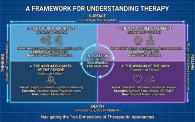
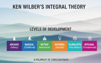
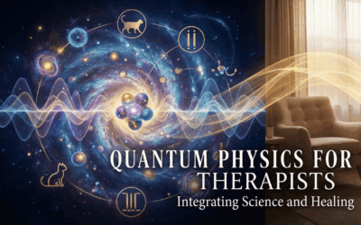
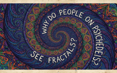
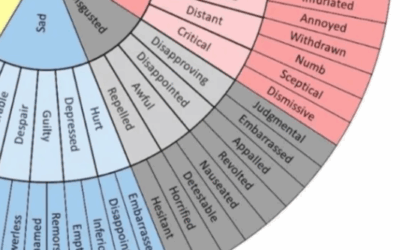


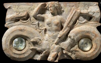
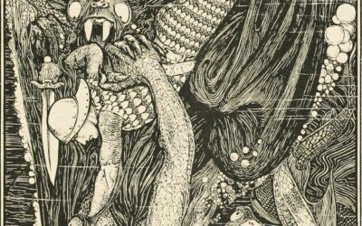


0 Comments